Download

1 / 43
430 likes | 461 Vues
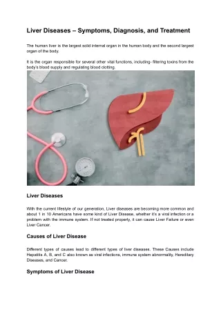
Learn how to detect, distinguish, and gauge liver damage with biochemical tests. Understand the significance of tests like bilirubin, aminotransferases, and more in liver disease evaluation and management.

E N D
Evaluation of Liver Function Dr. mousavi Abadan Khordad 1379
Several biochemical tests are useful in the evaluation and management of patients with hepatic dysfunction. • (1) detect the presence of liver disease • (2) distinguish among different types of liver disorders • (3) gauge the extent of known liver damage • (4) follow the response to treatment.
Liver test,They can be normal in patients with serious liver disease and abnormal in patients with diseases that do not affect the liver. They suggest a general category of liver disease, such as hepatocellular or cholestatic, which then further directs the evaluation.
Tests usually employed in clinical practice include: • The bilirubin • Aminotransferases • Alkaline phosphatase • Albumin • Prothrombin time When more than one of these tests provide abnormal findings or the findings are persistently abnormal on serial determinations, the probability of liver disease is high. When all test results are normal, the probability of missing occult liver disease is low.
TESTS BASED ON DETOXIFICATION AND EXCRETORY FUNCTIONS Serum Bilirubin: When measured by modifications of the original van den Bergh method, normal values of total serum bilirubin are reported between 1 and 1.5 mg/dLwith 95% of a normal population falling between 0.2 and 0.9 mg/dL. If the direct-acting fraction is less than 15% of the total, the bilirubin can be considered to all be indirect.
Elevation of the unconjugated fraction of bilirubin is rarely due to liver disease. • An isolated elevation of unconjugated bilirubin is seen primarily in hemolytic disorders and in a number of genetic conditions such as Crigler-Najjar and Gilbert’s syndromes • In the absence of hemolysis, an isolated, unconjugated hyperbilirubinemia in an otherwise healthy patient can be attributed to Gilbert’s syndrome, and no further evaluation is required.
In contrast, conjugated hyperbilirubinemia almost always implies liver or biliary tract disease. • The rate-limiting step in bilirubin metabolism is not conjugation of bilirubin, but rather the transport of conjugated bilirubin into the bile canaliculi. • Thus, elevation of the conjugated fraction may be seen in any type of liver disease. • In most liver diseases, both conjugated and unconjugated fractions of the bilirubin tend to be elevated.
In viral hepatitis, the higher the serum bilirubin, the greater is the hepatocellular damage. • Total serum bilirubin correlates with poor outcomes in alcoholic hepatitis. • It is also a critical component of the Model for End-Stage Liver Disease (MELD) score, a tool used to estimate survival of patients with end-stage liver disease and assess operative risk of patients with cirrhosis. • An elevated total serum bilirubin in patients with drug-induced liver disease indicates more severe injury.
Urine Bilirubin: • Unconjugated bilirubin always binds to albumin in the serum and is not filtered by the kidney. Therefore, any bilirubin found in the urine is conjugated bilirubin; the presence of bilirubinuria implies the presence of liver disease. • A urine dipstick test can theoretically give the same information as fractionation of the serum bilirubin. • This test is almost 100% accurate. Phenothiazines may give a false-positive reading with the Ictotest tablet. • In patients recovering from jaundice, the urine bilirubin clears prior to the serum bilirubin.
Blood Ammonia: Ammonia is produced in the body during normal protein metabolism and by intestinal bacteria, primarily those in the colon. • The liver plays a role in the detoxification of ammonia by converting it to urea, which is excreted by the kidneys. • Striated muscle also plays a role in detoxification of ammonia, where it is combined with glutamic acid to form glutamine. • Patients with advanced liver disease typically have significant muscle wasting, which likely contributes to hyperammonemia in these patients. • There is very poor correlation between either the presence or the severity of acute encephalopathy and elevation of blood ammonia. • It can be occasionally useful for identifying occult liver disease in patients with mental status changes.
The ammonia can be elevated in patients with severe portal hypertension and portal blood shunting around the liver even in the presence of normal or near-normal hepatic function. • Elevated arterial ammonia levels have been shown to correlate with outcome in fulminant hepatic failure.
Serum Enzymes : • The elevation of a given enzyme activity in the serum is thought to primarily reflect its increased rate of entrance into serum from damaged liver cells. Serum enzyme tests can be grouped into three categories: • (1) Enzymes whose elevation in serum reflects damage to hepatocytes • (2) Enzymes whose elevation in serum reflects cholestasis • (3) Enzyme tests that do not fit precisely into either pattern.
ENZYMES THAT REFLECT DAMAGE TO HEPATOCYTES They include aspartate aminotransferase (AST) and alanine aminotransferase (ALT). • AST is found in the liver, cardiac muscle, skeletal muscle, kidneys, brain, pancreas, lungs, leukocytes, and erythrocytes in decreasing order of concentration. • ALT is found primarily in the liver and is therefore a more specific indicator of liver injury.
Liver cell necrosis is not required for the release of the aminotransferases, and there is a poor correlation between the degree of liver cell damage and the level of the aminotransferases. Thus, the absolute elevation of the aminotransferases is of no prognostic significance in acute hepatocellular disorders. • The normal range for aminotransferases varies widely among laboratories, but generally ranges from 10–40 IU/L.
Levels of up to 300 IU/L are nonspecific and may be found in any type of liver disorder. • Minimal ALT elevations in asymptomatic blood donors rarely indicate severe liver disease; studies have shown that fatty liver disease is the most likely explanation. • Striking elevations—i.e., aminotransferases >1000 IU/L—occur almost exclusively in disorders associated with extensive hepatocellular injury such as: • (1) Viral hepatitis • (2) Ischemic liver injury (prolonged hypotension or acute heart failure) • (3) Toxin- or drug-induced liver injury.
The pattern of the aminotransferase elevation can be helpful diagnostically. • In most acute hepatocellular disorders, the ALT is higher than or equal to the AST. • Whereas the AST:ALT ratio is typically <1 in patients with chronic viral hepatitis and nonalcoholic fatty liver disease, • A number of groups have noted that as cirrhosis develops, this ratio rises to >1. An AST:ALT ratio >2:1 is suggestive, whereas a ratio >3:1 is highly suggestive, of alcoholic liver disease. • The AST in alcoholic liver disease is rarely >300 IU/L, and the ALT is often normal. • A low level of ALT in the serum is due to an alcohol-induced deficiency of pyridoxal phosphate.
The aminotransferases are usually not greatly elevated in obstructive jaundice. • One notable exception occurs during the acute phase of biliary obstruction caused by the passage of a gallstone into the common bile duct. In this setting, the aminotransferases can briefly be in the 1000–2000 IU/L range. However, aminotransferase levels decrease quickly, and the liver function tests rapidly evolve into those typical of cholestasis.
ENZYMES THAT REFLECT CHOLESTASIS • The activities of three enzymes: Alkaline phosphatase, 5′-nucleotidase, and γ-glutamyltranspeptidase (GGT)—are usually elevated in cholestasis. • Reflecting its more diffuse localization in the liver, GGT elevation in serum is less specific for cholestasis than are elevations of alkaline phosphatase or 5′-nucleotidase • Some have advocated the use of GGT to identify patients with occult alcohol use.
The normal serum alkaline phosphatase consists of many distinct isoenzymesfound in the liver; bone; placenta; and, less commonly, small intestine. Others condition with increase alkaline phosphatase: • Patients over age 60 (1–1.5 times normal) • Blood types O and B can have an elevation of the serum alkaline phosphatase after eating a fatty meal due to the influx of intestinal alkaline phosphatase into the blood. • Nonpathologically elevated in children and adolescents undergoing rapid bone growth because of bone alkaline phosphatase • late in normal pregnancies due to the influx of placental alkaline phosphatase.
Elevation of liver-derived alkaline phosphatase is not totally specific for cholestasis, and a less than threefold elevation can be seen in almost any type of liver disease. • Alkaline phosphatase elevations greater than four times normal occur primarily in patients with cholestatic liver disorders, Infiltrative liver diseases such as[ cancer and amyloidosis, and bone conditions characterized by rapid bone turnover (e.g., Paget’s disease)].
To distinguish liver alkaline phosphatase from others conditions: • First, and most precise, is the fractionation of the alkaline phosphatase by electrophoresis. • The second, best substantiated, and most available approach involves the measurement of serum 5′-nucleotidase or GGT.
In the absence of jaundice or elevated aminotransferases, an elevated alkaline phosphatase of liver origin often, but not always, suggests early cholestasis and, less often, hepatic infiltration by tumor or granulomata. Other conditions that cause isolated elevations of the alkaline phosphatase include : • Hodgkin’s disease • Diabetes • Hyperthyroidism • Congestive heart failure • Amyloidosis • Inflammatory bowel disease.
TESTS THAT MEASURE BIOSYNTHETIC FUNCTION OF THE LIVER Serum Albumin • Serum albumin is synthesized exclusively by hepatocytes. Serum albumin has a long half-life: 18–20 days, with ~4% degraded per day. • Not a good indicator of acute or mild hepatic dysfunction. • In hepatitis, albumin levels <3 g/dL should raise the possibility of chronic liver disease.
One exception is the patient with ascitesin whom synthesis may be normal or even increased, but levels are low because of the increased volume of distribution. Others coditions with hypoalbuminemia: In protein malnutrition of any cause Protein-losing enteropathies Nephrotic syndrome Chronic infections
Serum Globulins • Serum globulins are a group of proteins made up of γ globulins (immunoglobulins) produced by B lymphocytes and α and β globulins produced primarily in hepatocytes. • γ globulins are increased in chronic liver disease, such as chronic hepatitis and cirrhosis. • In cirrhosis, the increased serum γ globulin concentration is due to the increased synthesis of antibodies, some of which are directed against intestinal bacteria. • This occurs because the cirrhotic liver fails to clear bacterial antigens that normally reach the liver through the hepatic circulation.
Increases in the concentration of specific isotypes of γ globulins are often helpful in the recognition of certain chronic liver diseases. • Diffuse polyclonal increases in IgG levels are common in autoimmune hepatitis; increases >100% should alert the clinician to this possibility. • Increases in the IgM levels are common in primary biliary cholangitis. • whereas increases in the IgA levels occur in alcoholic liver disease.
COAGULATION FACTORS • With the exception of factor VIII, which is produced by vascular endothelial cells, the blood clotting factors are made exclusively in hepatocytes. • Their serum half-lives are much shorter than albumin, ranging from 6 h for factor VII to 5 days for fibrinogen. Helpful in both diagnosis and assessing the prognosis of acute parenchymal liver disease.
Useful for this purpose is the serum prothrombin time, which collectively measures factors II, V, VII, and X. • Biosynthesis of factors II, VII, IX, and × depends on vitamin K. • The international normalized ratio (INR) is used to express the degree of anticoagulation on warfarin therapy.
The prothrombin time may be elevated: • In hepatitis • Cirrhosis • Disorders that lead to vitamin K deficiency such as obstructive jaundice or fat malabsorption of any kind. Marked prolongation of the prothrombin time, >5 s above control and not corrected by parenteral vitamin K administration, is a poor prognostic sign in acute viral hepatitis and other acute and chronic liver diseases. The INR, along with the total serum bilirubin and creatinine, are components of the MELD score, which is used as a measure of hepatic decompensation and to allocate organs for liver transplantation.
OTHER DIAGNOSTIC TESTS • Although tests may direct the physician to a category of liver disease, additional radiologic testing and procedures are often necessary to make the proper diagnosis, as shown in Figure 358-1.
Percutaneous Liver Biopsy Liver biopsy is of proven value in the following situations: • (1) hepatocellular disease of uncertain cause • (2) prolonged hepatitis with the possibility of autoimmune hepatitis • (3) unexplained hepatomegaly • (4) unexplained splenomegaly • (5) hepatic filling defects by radiologic imaging • (6) fever of unknown origin • (7) staging of malignant lymphoma. Liver biopsy is most accurate in disorders causing diffuse changes throughout the liver and is subject to sampling error in focal infiltrative disorders such as hepatic metastases.
Contraindications to performing a percutaneous liver biopsy include: • Significant ascites • Prolonged INR. Under these circumstances, the biopsy can be performed via the transjugular approach.
Noninvasive Tests to Detect Hepatic Fibrosis FibroTest(marketed as FibroSure in the United States): • The test incorporates haptoglobin, bilirubin, GGT, apolipoprotein A-I, and α2-macroglobulin and has been found to have high positive and negative predictive values for diagnosing advanced fibrosis in patients with chronic hepatitis C, chronic hepatitis B, and alcoholic liver disease and patients taking methotrexate for psoriasis. Transient elastography(TE), marketed as FibroScan, and magnetic resonance elastography (MRE).
TE uses ultrasound waves to measure hepatic stiffness noninvasively. As such: • Chronic hepatitis C • Primary biliary cholangitis • Hemochromatosis • Nonalcoholic fatty liver disease • Recurrent chronic hepatitis after liver transplantation. MRE has been found to be superior to TE for staging liver fibrosis in patients with a variety of chronic liver diseases, but requires access to a magnetic resonance imaging scanner.
Ultrasonography • Ultrasonography is the first diagnostic test to use in patients whose liver tests suggest cholestasis, to look for the presence of a dilated intrahepatic or extrahepatic biliary tree or to identify gallstones. • In addition, it shows space-occupying lesions within the liver, enables the clinician to distinguish between cystic and solid masses, and helps direct percutaneous biopsies. Ultrasound with Doppler imaging can detect • the patency of the portal vein • hepatic artery • hepatic veins • determine the direction of blood flow. • This is the first test ordered in patients suspected of having Budd-Chiari syndrome.
Home message • A battery of tests that includes the aminotransferases, alkaline phosphatase, bilirubin, albumin, and prothrombin time along with the judicious use of the other tests described in this chapter. • It is often necessary to repeat these tests on several occasions over days to weeksfor a diagnostic pattern to emerge.