Energy and Cellular Metabolism
560 likes | 580 Vues
Explore the world of energy in biological systems, from chemical reactions to ATP production. Learn about enzymes, metabolic pathways, and the dynamic process of energy transfer and storage. Dive into the laws of thermodynamics and the intricate mechanisms of ATP production in cellular metabolism.

Energy and Cellular Metabolism
E N D
Presentation Transcript
Energy and Cellular Metabolism 4
About this Chapter • Energy in biological systems • Chemical reactions • Enzymes • Metabolism • ATP production • Synthetic pathways
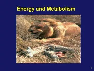
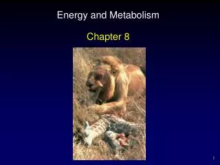
Energy: Biological Systems Energy transfer in the environment Figure 4-1
Energy: Capacity to Do Work • Chemical work • Making and breaking of chemical bonds • Transport work • Moving ions, molecules, and larger particles • Creates concentration gradients • Mechanical work • Used for movement
Energy: Two Forms The relationship between kinetic energy and potential energy Figure 4-2a
Energy: Two Forms Figure 4-2b
Energy: Two Forms Figure 4-2c
Energy: Thermodynamics • First law of thermodynamics • Total amount of energy in the universe is constant • Second law of thermodynamics • Processes move from state of order to disorder or entropy
Chemical Reactions: Overview Activation energy is the energy that must be put into reactants before a reaction can proceed A + B C + D Figure 4-3
Chemical Reactions: Coupling Energy transfer and storage in biological reactions Figure 4-5
Enzymes: Overview • Speed up the rate of reactions • Isozymes • Catalyze same reaction but under different conditions • May be activated, inactivated, or modulated • Coenzymes vitamins • Chemical modulators temperature and pH
Enzymes: Speed Up Reactions Enzymes lower the activation energy of reactions Figure 4-8
Enzymes: Law of Mass Action Figure 4-9a
Enzymes: Law of Mass Action Figure 4-9b
Metabolism: Overview A group of metabolic pathways resembles a road map Figure 4-10
Metabolism: Cell Regulation • Controlling enzyme concentrations • Producing modulators • Feedback inhibition • Using different enzymes • Isolating enzymes • Maintaining ratio of ATP to ADP • ADP + Pi + energy ATP
ATP Production: Overview Overview of aerobic pathways for ATP production Figure 4-13
ATP Production: Glycolysis Glucose + 2 NAD++ 2 ADP + P 2 Pyruvate + 2 ATP + 2 NADH + 2 H++ 2 H20 Figure 4-14
ATP Production: Pyruvate Metabolism Pyruvate can be converted into lactate or acetyl CoA Figure 4-15
ATP Production: Citric Acid Cycle • Acetyl CoA enters the citric acid cycle producing 3 NADH, 1 FADH2, and 1 ATP Figure 4-16
ATP Production: Electron Transport Mitochondrial matrix CITRIC ACID CYCLE Inner mitochondrial membrane O2 + Matrix pool of H+ 2 H2O 3 e– 1 ATP ADP + Pi 4e– 4 High-energy electrons ATP synthase H+ H+ H+ 2 Inter- membrane space H+ High-energy electrons Electron transport system H+ H+ H+ Outer mitochondrial membrane High-energy electrons from glycolysis Cytosol 3 4 1 2 Potential energy captured in the H+ concentration gradient is converted to kinetic energy when H+ pass through the ATP synthase. Some of the kinetic energy is captured as ATP. Energy released during metabolism is captured by high- energy electrons carried by NADH and FADH2. Electrons at the end of the electron transport system are back to their normal energy state. They combine with H+ and oxygen to form water. Energy from high-energy electrons moving along the protein complexes of the electron transport system pumps H+ from the matrix into the intermembrane space. Figure 4-17
ATP Production: Electron Transport Mitochondrial matrix CITRIC ACID CYCLE Inner mitochondrial membrane e– 1 High-energy electrons Inter- membrane space High-energy electrons Electron transport system Outer mitochondrial membrane High-energy electrons from glycolysis Cytosol 1 Energy released during metabolism is captured by high- energy electrons carried by NADH and FADH2. Figure 4-17, step 1
ATP Production: Electron Transport Mitochondrial matrix CITRIC ACID CYCLE Inner mitochondrial membrane e– 1 e– High-energy electrons H+ H+ H+ 2 Inter- membrane space High-energy electrons Electron transport system H+ H+ H+ Outer mitochondrial membrane High-energy electrons from glycolysis Cytosol 1 2 Energy released during metabolism is captured by high- energy electrons carried by NADH and FADH2. Energy from high-energy electrons moving along the protein complexes of the electron transport system pumps H+ from the matrix into the intermembrane space. Figure 4-17, steps 1–2
ATP Production: Electron Transport Mitochondrial matrix CITRIC ACID CYCLE Inner mitochondrial membrane O2 + Matrix pool of H+ 2 H2O 3 e– 1 4e– High-energy electrons H+ H+ H+ 2 Inter- membrane space High-energy electrons Electron transport system H+ H+ H+ Outer mitochondrial membrane High-energy electrons from glycolysis Cytosol 3 1 2 Energy released during metabolism is captured by high- energy electrons carried by NADH and FADH2. Energy from high-energy electrons moving along the protein complexes of the electron transport system pumps H+ from the matrix into the intermembrane space. Electrons at the end of the electron transport system are back to their normal energy state. They combine with H+ and oxygen to form water. Figure 4-17, steps 1–3
ATP Production: Electron Transport Mitochondrial matrix CITRIC ACID CYCLE Inner mitochondrial membrane O2 + Matrix pool of H+ 2 H2O 3 e– 1 ATP ADP + Pi 4e– 4 High-energy electrons ATP synthase H+ H+ H+ 2 Inter- membrane space H+ High-energy electrons Electron transport system H+ H+ H+ Outer mitochondrial membrane High-energy electrons from glycolysis Cytosol 3 4 1 2 Potential energy captured in the H+ concentration gradient is converted to kinetic energy when H+ pass through the ATP synthase. Some of the kinetic energy is captured as ATP. Energy released during metabolism is captured by high- energy electrons carried by NADH and FADH2. Energy from high-energy electrons moving along the protein complexes of the electron transport system pumps H+ from the matrix into the intermembrane space. Electrons at the end of the electron transport system are back to their normal energy state. They combine with H+ and oxygen to form water. NADH and FADH2 ATP by oxidative phosphorylation Figure 4-17, steps 1–4
ATP Production: Large Biomolecules • Glycogenolysis • Glycogen • Storage form of glucose in liver and skeletal muscle • Converted to glucose or glucose 6-phosphate
ATP Production: Large Biomolecules • Protein catabolism and deamination • Catabolism • Hydrolysis of peptide bonds • Deamination • Removal of amino group
ATP Production: Lipolysis If acetyl CoA production exceeds capacity for metabolism, production of ketone bodies results Figure 4-20
Synthesis: Gluconeogenesis Glucose can be made from glycerol or amino acids in liver and kidney Figure 4-21
Synthesis: Lipids Figure 4-22
Synthesis: Lipids Figure 4-22 (1 of 3)
Synthesis: Lipids Figure 4-22 (2 of 3)
Synthesis: Lipids Figure 4-22 (3 of 3)
Synthesis: Lipids Figure 4-22
Synthesis: Protein Gene Regulatory proteins 1 GENE ACTIVATION Constitutively active Regulated activity Induction Repression TRANSCRIPTION 2 mRNA siRNA 3 mRNA PROCESSING Alternative splicing Interference mRNA “silenced” Processed mRNA Nucleus • rRNA in ribosomes • tRNA • Amino acids Cytoplasm 4 TRANSLATION Protein chain 5 POST-TRANSLATIONAL MODIFICATION Cleavage into smaller peptides Addition of groups: • sugars • lipids • —CH3 • phosphate Folding and cross-links Assembly into polymeric proteins The major steps required to convert the genetic code of DNA into a functional protein 20 different amino acids made from 4 nitrogenous bases Figure 4-24
Synthesis: Protein Gene Regulatory proteins 1 GENE ACTIVATION Constitutively active Regulated activity Induction Repression Nucleus Cytoplasm Figure 4-24, step 1
Synthesis: Protein Gene Regulatory proteins 1 GENE ACTIVATION Constitutively active Regulated activity Induction Repression 2 TRANSCRIPTION mRNA Nucleus Cytoplasm Figure 4-24, steps 1–2
Synthesis: Protein Gene Regulatory proteins 1 GENE ACTIVATION Constitutively active Regulated activity Induction Repression 2 TRANSCRIPTION mRNA siRNA 3 mRNA PROCESSING Alternative splicing Interference mRNA “silenced” Processed mRNA Nucleus Cytoplasm Figure 4-24, steps 1–3
Synthesis: Protein Gene Regulatory proteins 1 GENE ACTIVATION Constitutively active Regulated activity Induction Repression 2 TRANSCRIPTION mRNA siRNA 3 mRNA PROCESSING Alternative splicing Interference mRNA “silenced” Processed mRNA Nucleus • rRNA in ribosomes • tRNA • Amino acids Cytoplasm 4 TRANSLATION Protein chain Figure 4-24, steps 1–4
Synthesis: Protein Gene Regulatory proteins 1 GENE ACTIVATION Constitutively active Regulated activity Induction Repression 2 TRANSCRIPTION mRNA siRNA 3 mRNA PROCESSING Alternative splicing Interference mRNA “silenced” Processed mRNA Nucleus • rRNA in ribosomes • tRNA • Amino acids Cytoplasm 4 TRANSLATION Protein chain 5 POST-TRANSLATIONAL MODIFICATION Addition of groups: • sugars • lipids • —CH3 • phosphate Cleavage into smaller peptides Folding and cross-links Assembly into polymeric proteins Figure 4-24, steps 1–5
Protein: Transcription • Transcription factors bind and activate promoter region • RNA polymerase binds and “unwinds” DNA • mRNA created from sense strand • mRNA is processed by • RNA interference • Alternative splicing
Protein: Transcription and Translation DNA 1 Transcription Nuclear membrane RNA polymerase mRNA processing 2 Amino acid Growing peptide chain tRNA Incoming tRNA bound to an amino acid 4 Translation Lys Asp Phe Trp Outgoing “empty” tRNA 3 Attachment of ribosomal subunits U U U C U A Anticodon A A C C G A A A G A A G U G U C U U mRNA Ribosome mRNA 5 Termination Each tRNA molecule attaches at one end to a specific amino acid. The anticodon of the tRNA molecule pairs with the appropriate codon on the mRNA, allowing amino acids to be linked in the order specified by the mRNA code. Ribosomal subunits Completed peptide Figure 4-27
Protein: Transcription and Translation DNA 1 Transcription Nuclear membrane RNA polymerase Figure 4-27, step 1
Protein: Transcription and Translation DNA 1 Transcription Nuclear membrane RNA polymerase mRNA processing 2 Figure 4-27, steps 1–2
Protein: Transcription and Translation DNA 1 Transcription Nuclear membrane RNA polymerase mRNA processing 2 3 Attachment of ribosomal subunits Figure 4-27, steps 1–3
Protein: Transcription and Translation DNA 1 Transcription Nuclear membrane RNA polymerase mRNA processing 2 Amino acid Growing peptide chain tRNA Incoming tRNA bound to an amino acid 4 Translation Lys Asp Phe Trp Outgoing “empty” tRNA 3 Attachment of ribosomal subunits U U U C U A Anticodon A A C C G A A A G A A G U G U C U U mRNA Ribosome Each tRNA molecule attaches at one end to a specific amino acid. The anticodon of the tRNA molecule pairs with the appropriate codon on the mRNA, allowing amino acids to be linked in the order specified by the mRNA code. Figure 4-27, steps 1–4
Protein: Transcription and Translation DNA 1 Transcription Nuclear membrane RNA polymerase mRNA processing 2 Amino acid Growing peptide chain tRNA Incoming tRNA bound to an amino acid 4 Translation Lys Asp Phe Trp Outgoing “empty” tRNA 3 Attachment of ribosomal subunits U U U C U A Anticodon A A C C G A A A G A A G U G U C U U mRNA Ribosome mRNA 5 Termination Each tRNA molecule attaches at one end to a specific amino acid. The anticodon of the tRNA molecule pairs with the appropriate codon on the mRNA, allowing amino acids to be linked in the order specified by the mRNA code. Ribosomal subunits Completed peptide Figure 4-27, steps 1–5
Protein: Post-Translational Modification • Protein folding • Creates tertiary structure • Cross-linkage • Strong covalent bonds disulfide • Cleavage • Addition of other molecules or groups • Assembly into polymeric proteins
Protein: Post-Translational Modification and the Secretory Pathway Figure 4-28