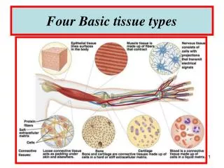
Four Basic tissue types
Four Basic tissue types. BLOOD. - Blood is sometimes considered to be a fluid connective tissue because of the mesenchymal origin of its cells and a low ratio of cells to liquid intercellular substance, the blood plasma .

Four Basic tissue types
E N D
Presentation Transcript
BLOOD -Blood is sometimes considered to be a fluid connective tissue because of the mesenchymal origin of its cells and a low ratio of cells to liquid intercellular substance, the blood plasma. - In human adults about 5 liter of blood contribute 7-8 % to the body weight of the individual. -The contribution of red blood cells (erythrocytes) to the total volume of the blood (haematocrit) is about 43%. -Erythrocytes are the dominant (99%) but not the only type of cells in the blood. - We also find leukocytes and, in addition, blood platelets. - Erythrocytes, leukocytes and blood platelets are also being referred to as the formed elements of the blood.. -Erythrocytes and blood platelets perform their functions exclusively in the blood stream. -In contrast, leukocytes reside only temporarily in the blood. -Leukocytes can leave the blood stream through the walls of capillaries and venules and enter either connective or lymphoid tissues.
Whole Blood Centrifuged with Anticoagulant Plasma (54%) Serum - plasma minus fibrinogen H2O -91-92% 6 3 Protein -7-8% -7-8% Red Cells - 5 X 10 / mm Electrolytes 3 3 White Cells Nutrients - 6-9 X 10 / mm Blood Gases Hormones Buffy Coat (1%) (white cells and platelets) Hematocrit (volume RBC’s/unit plasma) Red Cells (45%) (erythrocytes) males - 40-50% females- 35-40%
Red Blood Cells (erythrocytes) 7.5-8.5 um dia. Biconcave Discs Sickle Cells rapidly-flowing blood stagnant blood- rouleaux (valine-glutamine subst. b-hemoglobin chain) Erythrocyte Shape Varies with Tonicity Spherocytosis (spectrin alteration) hypotonic very hypotonic hypertonic isotonic (spheres) (ghosts)
Erythrocytes -Erythrocytes do not contain a nucleus. -They do contain haemoglobin, which fills almost the entire cytoplasm - Erythrocytes are unable to move actively, but they are remarkably elastic and can withstand deformation. -They are typically biconcave disks although their shape is influenced by osmotic forces. -The average diameter of the disk is ~7 µm. Since erythrocytes can be found in the vast majority of histological sections in small numbers even in perfused tissues , they will often allow us to estimate the size of other structures or cells. - Mature erythrocytes do not contain organelles, and their cytoplasm looks fairly homogenous even in the EM -At high magnification some granularity may be visible in EM images. -The granular appearance is caused by haemoglobin molecules - Foetal erythrocytes (up to the 4th month of gestation) are larger than "adult" erythrocytes, and they are nucleated. -The later feature they share with erythrocytes of other animal classes (e.g. amphibia and birds).
Functions of Erythrocytes -Erythrocytes function in the transport of oxygen. -Haemoglobin ; the oxygen binding protein in erythrocytes, contributes about 30% of the weight of an erythrocyte. -The lifespan of an erythrocyte in the bloodstream is 100-120 days -About 5×10¹¹ erythrocytes are formed/destroyed each day.
Microscopy of Erythrocytes Blood Smear- erythrocytes SEM- erythrocytes RED BLOOD CELLS
Leukocytes • Leukocytes can be further subdivided into : • granular leukocytes, i.e. neutrophils, basophils and eosinophils, • and non-granular leukocytes, i.e. monocytes and lymphocytes . -In healthy individuals the relative numbers of circulating leukocyte types are quite stable. -A differential leukocyte count would typically produce the following cell frequencies (numbers in parentheses are the range of normal frequencies reported in different texts) : • ~ 60% neutrophils (50% - 70%) • ~ 3% eosinophils (>0% - 5%) • ~ 0.5% basophils (>0% - 2%) • ~ 5% monocytes (1% - 9%) • ~ 30% lymphocytes (20% - 40%) • -Changes in their relative numbers indicate that something abnormal is happening in the organism. • -A larger than usual number of neutrophils (neutrophilia)would indicate e.g. an acute or chronic infection. • -The number of basophils and eosinophils may increase (eosinophilia or basophilia) as a consequence of e.g. allergic disorders.
Granular Leukocytes -Granular leukocytes are all approximately the same size about 12-15 µm in diameter. -Their nuclei form lobes, and nucleoli cannot be seen. -The number of nuclear lobes varies according to cell type. -All granulocytes are motile. -The term granulocytes refers to the presence of granules in the cytoplasm of these cells. -The granules correspond to secretory vesicles and lysosomes -Specific granulesare the granules which are only found in one particular type of granulocytes.
GRANULAR LEUKOCYTES Neutrophil (PMN) (55-65%) (polymorphonuclear leukocyte) Specific Granules (lysosomes & phagocytins) Lobes Azurophil Granules of (lysosomes) Nucleus Ectoplasmic Layer Neutrophil Specific Pseudopodia Granules
Neutrophil granulocytes (or neutrophils) -Have a very characteristic nucleus. -It is divided into 3-5 lobes which are connected by thin strands of chromatin. -The number of lobes increases with cell age. Up to 7 lobes can be found in very old neutrophils (hypersegmented cells). -Neutrophils (like all other granulocytes, monocytes and lymphocytes) contain all the organelles that make up a typical cell. -In addition to the usual complement of organelles, they also contain two types of granules : I) Primary granules (or A granules) contain lysosomal enzymes and are likely to be primary lysosomes, although they are larger (0.4 µm) than the "ordinary" primary lysosome. II) Secondary granules (or B granules), the specific granules of the neutrophils, contain enzymes with strong bactericidal actions. -The specific granules of neutrophils stain only weakly if they are at all visible they are "neutral", hence the term neutrophil.
Functions of neutrophils -Neutrophils play a central role in inflammatory processes. -Large numbers invade sites of infection in response to factors (e.g. cytokines) released by cells which reside at an infection site. -Neutrophils are the first wave of cells invading infection sires - Receptors in their plasma membrane allow them to recognise foreign bodies, e.g. bacteria, and tissue debris, which they begin to phagocytose and destroy. -The phagocytotic activity of neurophils is further stimulated if invading microorganisms are "tagged" with antibodies (or opsonised). -Neutrophils cannot replenish their store of granules. -The cells die once their supply of granules has been exhausted. -Dead neutrophils and tissue debris are the major components of pus -Their lifespan is only about one week. -Lost neutrophils are quickly replenished from a reserve population in the bone marrow. -Because they are younger, their nuclei have fewer lobes than the "average" neutrophil. - A high proportion of neutrophils, with few nuclear lobes indicates a recent surge in their release from the bone marrow.
Shown in the following photo are red blood cells in a smear, small platelets (p) and a neutrophil with a multilobed nucleus. The drumstick like projection from the nucleus is called a "Barr Body" and represents the condensed X chromosome. This signifies that the blood was taken from a female
GRANULAR LEUKOCYTES Eosinophils (1-3%) Eosinophilic (acidophilic) granules (destroy antigen-antibody complexes) Eosinophil Crystals Specific Granules
Eosinophil granulocytes (or eosinophils) - Their nucleus usually has only two lobes. - Almost all of the cytoplasm appears filled with the specific granules of the eosinophils. As the term "eosinophil" indicates, these granules are not neutral but stain red or pink when eosin or a similar dye is used in the staining process. - Aside from the usual complement of organelles eosinophils contain some large rounded vesicles (up to 1 µm) in their cytoplasm. - These granules correspond to the eosinophilic grains that we see in the light microscope. - The specific granules contain, in addition to enzymes that otherwise are found in lysosomes, an electron-dense, proteinaceous crystal. - This crystal is composed of major basic protein (MBP).
Functions of eosinophils granulocytes • -The presence of antibody-antigen complexes stimulates the immune system. • -Eosinophils phagocytose these complexes and this may prevent the immune system from "overreacting". • -Their granules also contain the enzymes histaminase and arylsulfatase. • -These enzymes break down histamine and leukotrienes, which again may dampen the effects of their release by basophils or mast cells. • -MBP, which can also function as a cytotoxin, and its release by eosinophils may be involved in the response of the body against parasitic infections, which are accompanied by an increase in the number of eosinophils.
Granular Leukocytes Basophils (less than 1%) Basophilic Granules heparin histamine serotonin Basophil
Basophil granulocytes (or basophils) -Basophilic granulocytes have a 2 or 3 lobed nucleus -The lobes are usually not as well defined as in neutrophilic granulocytes and the nucleus may appear S-shaped. -The specific granules of basophils are stained deeply bluish or reddish-violet. -Their colour corresponds closely to the colour of the nucleus which sometimes is difficult to see amongst or behind the granules. -The granules are not as numerous as those in eosinophils. -The specific granules of basophils (about 0.5 µm) appear quite dark in EM pictures. -They contain heparin, histamine lysosomal enzymes and leukotrienes(the later correspond to the slow-reacting substance of anaphylaxis or SRS-A).
Eosinophil, Neutrophil and Basophil Eosinophil Neutrophil Basophil
AGRANULAR LEUKOCYTES Monocytes (3-8%) Lymphocytes (20-25%) B Lymphocytes (humoral immunity) Mononuclear T-Lymphocytes Phagocytic System (cell mediated immunity) Monocytes Lymphocytes
Non-granular leukocytes Monocytes -These cells can be slightly larger than granulocytes (about 12-18 µm in diameter). -Their cytoplasm stains usually somewhat stronger than that of granulocytes, but it does not contain any structures which would be visible in the light microscope using most traditional stains (a few very fine bluish grains may be visible in some monocytes). -The "textbook" monocyte has a C-shaped nucleus. -Monocytes contain granules (visible in the EM) which in appearance and content correspond to the primary granules of neutrophils, i.e. the granules correspond to lysosomes.
Functions of monocytes - Once monocytes enter the connective tissue they differentiate into macrophages. - At sites of infection macrophages are the dominant cell type after the death of the invading neutrophils. - They phagocytose microorganisms, tissue debris and the dead neutrophils. - Monocytes also give rise to osteoclasts, which are able to dissolve bone. - They are of importance in bone remodelling
Lymphocytes -These cells are very variable in size. -The smallest may be smaller than erythrocytes (down to ~5 µm in diameter) while the largest may reach the size of large granulocytes (up to 15 µm in diameter). -How much cytoplasm is discernible depends very much on the size of the lymphocyte. -In small ones, which are the majority of lymphocytes in the blood, the nucleus may appear to fill the entire cell. -Large lymphocytes have a wider rim of cytoplasm which surrounds the nucleus. -Both the nucleus and the cytoplasm stain blue (and darker than most other cell types in the blood). -The typical lymphocyte only contains the usual complement of cellular organelles. -The appearance of lymphocytes may change drastically when they are activated .
Functions of lymphocyte - Most lymphocytes in the blood stream belong to either the group of B-lymphocytes (~5%) or the group of T-lymphocytes (~90%). Unless they become activated, the two groups can not easily be distinguished using routine L or E/M. - Upon exposure to antigens by antigen-presenting cells (e.g. macrophages) and T-helper cells (one special group of T-lymphocytes) B-lymphocytes differentiate into antibody producing plasma cells . The amount of cytoplasm increases and RER fills a large portion of the cytoplasm of plasma cells. - T-lymphocytes represent the "cellular arm" of the immune response (cytotoxic T cells) and may attack foreign cells, cancer cells and cells infected by e.g. a virus. - T-lymphocytes and B-lymphocytes form the vast majority of lymphocytes in the blood stream, but they do not add up to 100%, and they usually are small lymphocytes. - The much less frequent medium-sized or large lymphocytes may represent e.g. natural killer ( Nk -) cells which belong to the group of large granular lymphocytes, or haemopoietic stem cells of which a few will be circulating in the blood stream.
BLOOD PLATELETS (thrombocytes) microtubules micro- tubules granules glycogen Hyalomere (pale blue) Granulomere (purple granules) glycogen Platelet- light micrograph microtubules microtubules
Blood Platelets (or thrombocytes) - Blood platelets do not contain a nucleus , unlike erythrocytes, which also lack a nucleus, the blood platelets of mammals have never been nucleated cells. - Instead, blood platelets are fragments of the cytoplasm of very large thrombocyte precursor cells, megakaryocytes. Like other cells involved in the formation in blood cells, megakaryocytes are found in the bone marrow. - Platelets are about 3 µm long but appear somewhat smaller in the microscope. - This is because their cytoplasm is divided into two zones: an outer hyalomere, which hardly stains, and an inner granulomere, which contains bluish staining granules. - These granules are usually not individually visible with the highest magnification on your microscope, and the granulomere appears more or less homogeneously blue. - In addition to different types of vesicles (i.e. the granules), mitochondria, ribosomes, lysosomes and a little ER are present in the thrombocyte granulomere. - Different types of vesicles contain either serotonin (electron-dense delta granules; few) or compounds important for blood coagulation(alpha granules - they also contain platelet-derived growth factor (PDGF) which may play a role in the repair of damaged tissue). - The hyalomere contains cytoskeletal fibres, which include actin and myosin.
Function of Blood Platelets - Platelets assist in haemostasis, the arrest of bleeding - Serotonin is a potent vasoconstrictor - The release of serotonin from thrombocytes, which adhere to the walls of a damaged vessels, is sufficient to close even small arteries - Platelets, which come into contact with collagenous fibers in the walls of the vessel (which are not usually exposed to the blood stream), swell, become "sticky" and activate other platelets to undergo the same transformation. This cascade of events results in the formation of a platelet plug (or platelet thrombus). - Finally, activating substances are released from the damaged vessel walls and from the platelets. These substances mediate the conversion of the plasma protein prothrombin into thrombin. - Thrombin catalyzes the conversion of fibrinogen into fibrin, which polymerizes into fibrils and forms a fibrous net in the arising blood clot. - Platelets captured in the fibrin net contract leading to clot retraction, which further assists in haemostasis. - Blood coagulation is a fairly complex process, which involves a large number of other proteins and messenger substances. Deficiencies in any one of them, either inherited or acquired, will lead to an impairment of haemostasis.
Erythrocytes, Lymphocytes, Neutrophils and Platelets Bar Body
Blood Clotting (released from platelets and injured Prothrombin Thromboplastin vessel wall) Thrombin Fibrin (clot) Fibrinogen (feltwork of fibrils, platelets and blood cells) thrombus platelets lesion
Blood Plasma Plasma- transparent, yellow liquid vehicle for cells Serum- plasma without fibrinogen Serum Proteins: Albumincolloid osmotic pressure and provide for solubility of hydrophobic components (e.i. lipids) Globulins gamma globulins- e.i. IgG (immune globulins-antibodies) beta globulins - hormone transport (e.i. thyrogloblin) Transferrin- iron transport Cerruloplasmin- copper transport Serum Lipoproteins: -chylomicrons- fatty acid transport -Very Low Density Lipoproteins (VLDL)) -Low Density Lipoproteins (LDL) -High Density Lipoproteins (HDL)
Haemopoiesis Where blood is made - Haemopoietic cells (those which produce blood) first appear in the yolk sac of the 2-week embryo. - By 8 weeks, blood making has become established in the liver of the embryo, and - by 12-16 weeksthe liver has become the major site of blood cell formation. It remains an active haemopoietic site until a few weeks before birth. The spleen is also active during this period, particularly in the production of lymphoid cells, and the foetal thymus is a transient site for some lymphocytes. The highly cellular bone marrow becomes an active blood making site from about 20 weeksgestation and gradually increases its activity until it becomes the major site of production about 10 weeks later. - At birth, active blood making red marrow occupies the entire capacity of the bones and continues to do so for the first 2-3 years after birth. The red marrow is then very gradually replaced by inactive, fatty, yellow, lymphoid marrow. The latter begins to develop in the shafts of the long bones and continues until, by 20-22 years, red marrow is present only in the upper ends of the femur and humerus and in the flat bones of the sternum, ribs, cranium, pelvis and vertebrae. However, because of the growth in body and bone size that has occurred during this period, the total amount of active red marrow (approximately 1000-1500 g) is nearly identical in the child and the adult.
- Adult red marrowhas a large reserve capacity for cell production. In childhood and adulthood, it is possible for blood making sites outside marrow, such as the liver, to become active if there is excessive demand as, for example, in severe haemolytic anaemia or following haemorrhage. - In old age, red marrow sites are slowly replaced with yellow, inactive marrow. -Red marrow forms all types of blood cell and is also active in the destruction of red blood cells. -Red marrow is, therefore, one of the largest and most active organs of the human body, approaching the size of the liver in overall mass although as mentioned it is distributed in various parts of the body. -About two-thirds of its mass functions in white cell production (leucopoiesis), and one-third in red cell production (erythropoiesis). However as we have already seen there are approximately 700 times as many red cells as white cells in peripheral blood. This apparent anomaly reflects the shorter life span and hence greater turnover of the white blood cells in comparison with the red blood cells.
Bone and bone marrow • Bone marrow is a spongy material that is found inside the bones (particularly the pelvic bones). • Stem cells are blood cells at the earliest stage of development in the bone marrow. • Within the bone marrow, stem cells develop into the different blood cells described below. • When the cells are fully mature they are released into the bloodstream. • Like a factory, bone marrow produces the cells which develop into the three different types of blood cells: • red blood cells, white blood cells, platelets, . • Normally, most of the stem cells in the body are in the bone marrow and there are only very small numbers in the bloodstream. However, it is possible to stimulate the stem cells to move into the bloodstream, by using low doses of certain chemotherapy drugs or injections of growth factors. Stem cells can be collected from the bloodstream or from the bone marrow. • A transplant using stem cells collected from the bone marrow is sometimes called a bone marrow transplant, when in fact it is really a transplant of stem cells.
-Haemopoietic cells surround the vascular sinusoids and are supported by reticular connective tissue -In addition to the endothelial cells of the sinusoids and the reticulocytes of the connective tissue, macrophages are frequent in red bone marrow. Megakaryocyte Endothelial Cells Diapedesis Adipocyte Endothelial Discontinuity Blood Sinusoid Hemopoetic Reticular Compartment Fibers
Haemopoietic Cells • -The basis of haemopoiesis is a small population of self-replicating stem cells, which ultimately can generate all types of blood cells. • -Their progeny may develop into either lymphocytic stem cells or pluripotent haemal stem cells (colony-forming unit - stem cell - CFU-S). • -The latter type gives rise to stem cells which can form the major groups of blood cells other than lymphocytes. • -Depending on their progeny it is possible to differentiate burst-forming unit of the erythroid line (BFU-E), colony-forming unit - granulocytes and macrophages (CFU-G/M), and colony-forming unit - megakaryocytes (CFU-Mk).
ERYTHROPOEISIS - The first identifiable stage of erythropoiesis is the proerythroblast - a large, slightly basophilic cell, which contains a large, lightly stained nulceus. - Proerythroblasts proliferate to generate a sequence of cells which show a gradual decrease in size and condensation of their chromatin. - They are named after changes in the staining characteristic of their cytoplasm (basophilic erythroblast, polychromatophilic and orthochromic normoblasts). - The nucleus is finally extruded from the normoblast - The cell enters circulation as a reticulocyte, which still contains some organelles. - Reticulocytes remain for a few days in either the bone marrow or the spleen to mature to erythrocytes.
ERYTHROPOEISIS Polychroma- Orthochroma- Nuclear Proerythroblast Basophilic Reticulocyte Hemocytoblast tophilic tophilic Extrusion Erythroblast RBC Erythroblast Erythroblast Pluripotential 1. dispersed 1. clumped 1. condensed 1. condensed 1. anucleate eccentric Stem Cell chromatin chromatin chromatin 2. spherical nucleus 2. nucleoli 2. no 2. grey-green 3. slight 2. pink nucleoli cytoplasm 3. cytoplasmic basophilia cytoplasm 3. maximum (hemoglobin basophilia (free ribosomes) basophilia synthesis)
MYELOPOEISIS - Myeloblast appear light-microscopically similar to proerythroblast. - They proliferate to generate promyelocytes. - Promyelocytes begin to accumulate nonspecific granules, but they are still able to divide. - The maturation of their progeny, the myeocytes, is characterised by the accumulation of specific granules and changes in nuclear morphology. - Metamyelocytes have a C-shaped nucleus.
MYELOPOEISIS - ( granulopoeisis ) Hemocytoblast Myeloblast (multipotential) (multipotential) Promyelocyte (multipotential) 1. loss of cytoplasmic basophilia 2. dispersed chromatin Basophilopoeisis Eosinophilopoeisis Neutrophilopoeisis
EOSINOPHILOPOEISIS Eosinophilic Eosinophilic Eosinophil Metamyelocyte Myelocyte (band cell) Specific and Metachromatic Granules
NEUTROPHILOPOEISIS NEUTROPHILIC METAMYELOCYTE Neutrophil NEUTROPHILIC MYELOCYTE
BASOPHILOPOEISIS Basophilic Basophilic Basophil Myelocyte Metamyelocyte
THROMBOPOEISIS - Are, as mentioned above, fragments of the cytoplasm of megakaryocytes. - Megakaryocytes are very large cells (up to 160 µm in diameter), which contain very large, highly lobulated, polyploid nuclei. - Megakaryocytes are in turn the product of the differentiation of basophilic megakaryoblasts.
THROMBOPOEISIS Endomitosis (w/o karyokinesis Hemocytoblast (w/o cytokinesis) Reserve Megakaryocyte (megakaryoblast) Platelet-Forming Megakaryocyte Residual Megakaryocyte Platelets Platelet-Shedding (fusion of plasma membrane)
This diagram shows the formation of the different types of blood cells from a common source the stem cell