Brain Tumor
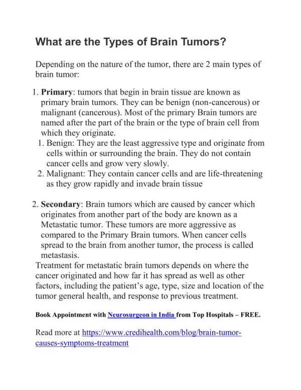
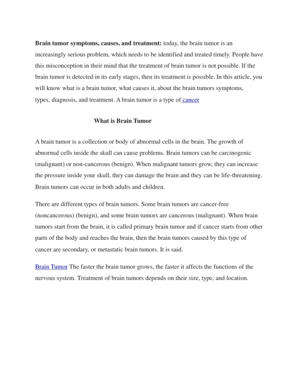
Brain Tumor. What is it?. Brain neoplasms are a diverse group of primary ( nonmetastatic ) tumors arising from one of the many different cell types within the central nervous system. Malignant brain tumors are defined by histopathologic features and a rapidly progressive pattern of growth

Brain Tumor
E N D
Presentation Transcript
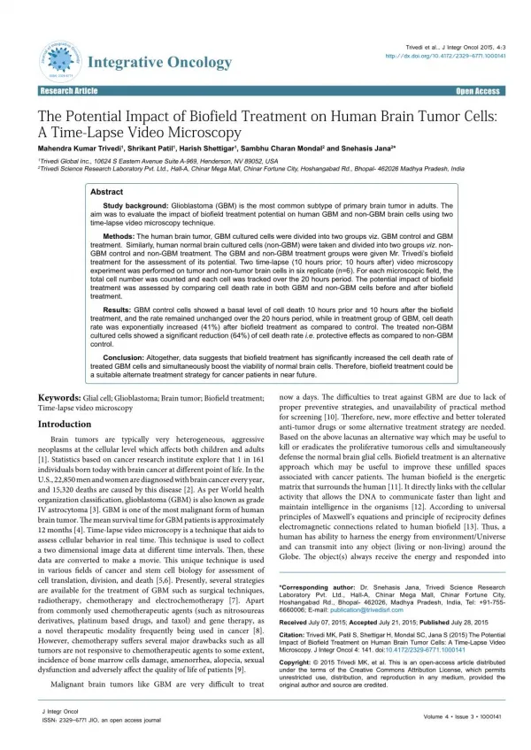
What is it? • Brain neoplasms are a diverse group of primary (nonmetastatic) tumors arising from one of the many different cell types within the central nervous system. • Malignant brain tumors are defined by histopathologic features and a rapidly progressive pattern of growth • Glioblastoma is the most common primary brain tumor in adults, accounting fo 50% to 60% of primary brain tumors
Pathophysiology • Nervous system tumors are clonal proliferations that develop secondary to changes in key growth regulatory genes • These genetic changes result in powerful growth advantages that enable the cells to priloferate, evolve, and disseminate • The etiologies of brain tumors remain unknown. • To date, only radiation and hereditary predisposition are clearly implicated as etiological factors • The WHO classification currently lists more than 100 types of nervous system tumors and their variants • This classification system allows for consideration of key clinical and imaging characteristics which can narrow the differential diagnosis to only a few common possibilities
Pathophysiology • Only about 5% of primary brain tumors have known hereditary factors • Li-Fraumeni syndrome, p53 defects, neurofibromatosis 1 (NF1) and 2 (NF2), tuberous sclerosis, von Hippel-Lindau disease, Turcot’s syndrome, and familial polyposis • Meningiomas • Strongest genetic link has been associated with NF2, an almost 50% incidence • Known to express estrogen and progesterone receptors. • High incidence of somatostatin receptors has also been found • Significance of these findings is uncertain but has led to diagnostic tests and treatment strategies • Radiation is only definite cause • Oligodendrogliomas • Viral infections (specifically the JC virus) has been implicated, but data are inconclusive
Incidence • Glioblastomas occur in approximately 2-3 cases/100,000 persons • Most commonly diagnosed primary brain tumor of adults is glioblastomamulitforme (grade IV) • Slightly more common in whites than blacks, Latinos, and Asians • Slightly more common in men than women with a male:female ration of 3:2 • Peak incidence is between 45 and 70 years. • Approximately 10% of glioblastomas occur in children
Risk Factors • Prior radiation may increase risk for primary brain tumor
Signs and Symptoms • Headache • Most common presenting complaint – reported by approximately 50% of the patients • Seizures (occur in 30%-60% of cases) • Signs and symptoms of hydrocephalus (headache, vomiting, clouding of consciousness, papilledema) • Memory loss • Focal motor weakness • Visual changes • Language deficits • Cognitive disturbances • Memory changes
Clinical Presentation • Worsening headache • Present for weeks to months • Classic triad of brain tumor headache: • Sleep disturbances • Severe pain • Nausea and vomiting • Headache is often bilateral and worsened by coughing, sneezing, bending, defecation, and sexual intercourse
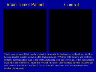
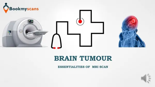
Imaging • MRI with and without contrast is the imaging study of choice • CT scanning is useful if calcification or hemorrhage is suspected • PET scan is helpful to distinguish neoplastic lesions (with high rate of metabolism) from other lesions such as demyelination or radiation necrosis (with a much lower metabolic rate)
Treatment • Surgery • Radiation • Palliative chemotherapy
References • Bradley W G. Neurology in Clinical Practice. 5th edition ed. Philadelphia: Butterworth Heinemann Elsevier; 2008. • Carey WD. Cleveland Clinic: Current Clinical Medicine. 2nd edition ed. Philadelphia: Saunders Elsevier; 2010. • Ferri F F. Ferri's Clinical Advisor 2011: Instant Diagnosis and Treatment. 1st ed. Philadelphia: Elsevier; 2011. • Marx J A. Rosen's Emergency Medicine. Vol 1. 7th edition ed. Philadelphia: Mosby Elsevier; 2010.