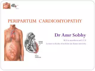
PERIPARTUM CARDIOMYOPATHY
420 likes | 804 Vues
PERIPARTUM CARDIOMYOPATHY. Dr.T.Venkatachalam Professor of Anaesthesiology Madras Medical College, Chennai. PERIPARTUM CARDIOMYOPATHY.

PERIPARTUM CARDIOMYOPATHY
E N D
Presentation Transcript
PERIPARTUM CARDIOMYOPATHY Dr.T.Venkatachalam Professor of Anaesthesiology Madras Medical College, Chennai
PERIPARTUM CARDIOMYOPATHY • Peripartum cardiomyopathy is defined as the onset of acute heart failure without demonstrable cause in the last trimester of pregnancy or within the first 5 months after delivery.
PERIPARTUM CARDIOMYOPATHY • Aform of Dilated Cardiomyopathy • Left ventricular systolic dysfunction • Results in signs and symptoms of heart failure • Often unrecognized, as symptoms of normal pregnancy commonly mimic those of mild heart failure.
Criteria for Peripartum Cardiomyopathy 1.Development of Cardiac failure in the last month of pregnancy or within 5 month after delivery 2. Absence of an identifiable cause for the cardiac failure. 3.Absence of recognizable heart disease prior to the last month of pregnancy. 4.Left ventricular systolic dysfunction demonstrated by classic Echo Cardio Graphic criteria such as depressed shortening fraction or ejection fraction. The National Heart, Lung and Blood Institute and the Office of rare diseases (1997)
Incidence • The incidence in the west ranges from 1 in 4000 deliveries • Sixty percent present within the first 2 months postpartum • Up to 7% may present in the last trimester of pregnancy. • Geographic variations exist with a higher incidence reported in areas of Africa because of malnutrition and local customs in the puerperium
Etiology Still unknown. • -nutritional deficiencies • -small vessel coronary artery abnormality • -hormonal effects • -toxemia • -maternal immunologic response to fetal antigen or • -myocarditis
Predisposing factors • -maternal age greater than 30 yr • -multiparous or eclamptic patients • - twinning • - racial origin (black) • - hypertension and • - nutritional deficiencies • In majority of cases there is no family history
Symptoms Symptoms of worsening cardiac failure like: • -dyspnoea on exertion • -fatigue • -ankle oedema • -embolic phenomena • -atypical chest pains and • -haemoptysis. • Many of above symptoms may occur even in normal pregnancy and can be mistaken for a diseased state.
Signs • -evidence of a raised CVP • -tachycardia • -cardiomegaly with a gallop rhythm (S3) • -mitral regurgitation • -pulmonary crackles and • -peripheral oedema.
PERIPARTUM CARDIOMYOPATHY On auscultation of the heart: • loud first heart sound • exaggerated splitting • mid systolic murmur and • continuous venous hum • These physical signs may confuse and there could be mistakes in the form of over diagnosis or disregarding of heart disease.
PERIPARTUM CARDIOMYOPATHY Chest radiograph: • cardiomegaly with pulmonary oedema • pulmonary venous congestion. The ElectroCardioGram: • nonspecific ST and T wave changes • atrial or ventricular arrhythmias and • conduction defects.
Echocardiography / Doppler • may reveal enlargement of all four chambers with marked reduction in left ventricular systolic function • small to moderate pericardial effusion and • mitral, tricuspid and pulmonary regurgitation • Ventricular wall motion, ejection fraction and cardiac output are decreased and • pulmonary wedge pressure is increased.
PERIPARTUM CARDIOMYOPATHY • The clinical presentation and hemodynamic features in PPCM are indistinguishable from those of other forms of dilated cardiomyopathy. • In the absence of any cardiac symptoms, one of the early indications about this condition is revealed during evaluation of the fetus with a fetal monitor and ultrasound
PERIPARTUM CARDIOMYOPATHY • Fetal growth is dependent on good blood flow to the uterus and placenta • An insufficient blood flow meansdecreased oxygenation resulting inslowed growth • This should prompt further investigation to discover heart disease.
The prognosis 50-60% patients show complete or near complete recovery within the first 6 months postpartum In others, either continued clinical deterioration leading to early death or persistent left ventricular dysfunction and chronic heart failure results There is an initial high risk period with mortality of 25-50% in the first 3 months postpartum. Patients with persistent cardiomegaly at 6 months have a reported mortality of 85% at 5 years.
The prognosis • Subsequent pregnancies in women with PPCM are often associated with relapses and high risk for maternal morbidity and mortality. • should be discouraged in women with PPCM who have persistent cardiac dysfunction.
Management of PPCM Vigorous treatment of acute heart failure. • Oxygen, diuretics, digoxin and vasodilators • Use of ACE inhibitors in early pregnancy should be avoided as it has teratogenic effects on fetus
PERIPARTUM CARDIOMYOPATHY • Anticoagulant therapy is recommended because of high incidence of thrombo embolic events in PPCM • Patient on oral anticoagulants require change to parenteral anticoagulants with short half life • Dose adjusted according to the PTT which may be discontinued before delivery. • After delivery Warfarin may be used
PERIPARTUM CARDIOMYOPATHY • Since the disease may be reversible, the temporary use of Intra Aortic Balloon Pump or LV assist device may help to stabilize the patient’s condition pending improvement.
PERIPARTUM CARDIOMYOPATHY • Many patients with PPCM show evidence of myocarditis in biopsy specimens. • Dobutamine stress echocardiography - for evaluating contractile reserve in women with recovered systolic function who are contemplating further pregnancies.
PERIPARTUM CARDIOMYOPATHY • Autopsy shows cardiac enlargement, often with mural thrombi along with histological evidence of myocardial degeneration and fibrosis.
The anaesthetic considerations Similar for any patient with heart failure presenting for caesarian section regardless of etiology • Hemodynamic goals include: • Maintenance of normal to low heart rate to decrease oxygen demand • Prevention of large swings in blood pressure. These goals can be achieved by giving either general or regional anesthesia
General anaesthesia • During GA important factors to keep in mind are: 1. Volatile agents that decrease LV contractility without dramatic vasodilatation is desirable. 2. Avoid agents that decease preload and after load. eg. hypovolemia, nitroglycerine, nitroprusside 3. Avoid agents that directly or indirectly increase heart rate and contractility like Pancuronium, atropine, epinephrine, ephedrine. .
General anaesthesia -cont 4. Replace Blood loss promptly. 5. Hypotension better treated with volume expansion and pure alpha adrenergic agonist. 6. Remember that insertion of CVP / PAC may induce atrial or ventricular dysarrhytmias
GA - Drawbacks • IV and Inhalational agents Cardiac depression • High dose Narcotics – Need for post op ventilation for both mother and child. -There is an increased risk of gastric aspiration. • The management of a failed intubation may become difficult by the longer acting nature of these drugs with mask ventilation and if associated with obesity.
Central Neuraxial Anaesthesia • The consideration for in these patients are similar to those with other causes of heart failure. • Subarachnoid block may better be avoided in these patients because of sudden onset of hemodynamic instability.
Central Neuraxial Anaesthesia Epidural anaesthesia -better choice • incremental doses • with opioids. May improve myocardial performance and the cardiac output by decreasing the systemic vascular resistance, thus reducing the after load on the left ventricle without impairing contractility
Central Neuraxial Anaesthesia • Pulmonary Artery catheter can guide fluid and inotrope requirements • Preloading to be avoided in these patients • Small bolus doses of 0.5% Bupivacaine or 2.0% Xylocaine (10 to 12 ml in L2 to L4) along with fentanyl up to 40 µg may be preferred.
Intra operative monitoring • Depends on the preoperative signs and symptoms. • In asymptomatic patients, a central venous catheter is adequate with non invasive BP monitoring. • In symptomatic patients or with echo findings of left ventricular dysfunction, a PA catheter and an arterial line if available will be useful.
PERIPARTUM CARDIOMYOPATHY • Oxytocin infusion is preferable • As infusion it will not produce sudden vasodilatation and hypotension. • It also helps to decrease the after load maintaining the hemodynamic stability
Post operative Care • It is better to monitor these patients in an ICU for hemodynamic stabilization. • It may worsen due to retention of water due to ant diuretic effect of Oxytocin • Re absorption of third space fluid after 48 hrs of the caesarian section. • The above factors increase the preload, worsening the patient’s condition.
Prognosis & Sequele • May develop a reduction in the left ventricular systolic function during subsequent pregnancies • This reduction would be greater in those with persistent left ventricular dysfunction at the start of the pregnancies. • Symptoms of heart disease develop in about 20% of women whose systolic function is normal at the start of the subsequent pregnancy and in almost half of the women who have persistent left ventricular dysfunction
Prognosis & Sequele • The out come is highly variable. • Some develop persistent disease while some return to normal state slowly. • These patients has a better survival rate than other types of cardiomyopathy.
REMEMBER • PPCM mimics changes occurring in normal pregnancy • Fetal growth retardation may point towards this condition • Treat like any other cardiac failure along with anti coagulant therapy • Epidural anaesthesia is preferable and continue monitoring in an ICU • Advice against subsequent pregnancies.
PERIPARTUM CARDIOMYOPATHY THANK YOU