Download
1 / 37
370 likes | 382 Vues
This text provides an overview of protein digestion and the metabolism of amino acids in the human body. It explains the process of protein breakdown and the role of various enzymes in digestion. It also discusses the different fates of amino acid degradation products and alternative methods of nitrogen excretion. Additionally, it covers the processing of amino acid carbon skeletons and their utilization in gluconeogenesis.

E N D
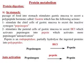
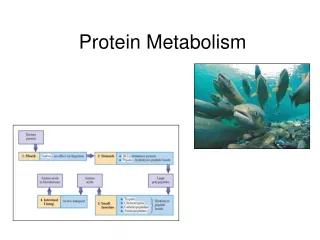
Protein Digestion Protein breakdown begins in the stomach. No protein hydrolyzing enzymes are found in saliva. Hydrolysis (10% of peptide bonds) & denaturization by pepsin enzyme & HCl acid produce short chain polypeptides in the stomach. Trypsin, chymotrypsin, & carboxypeptidase from Pancreatic juices, and Aminopeptidase from cells in the small intestine Brush Zone create “free” amino acids.
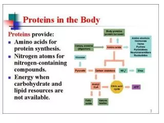
Free amino acids are absorbed thru intestinal wall via active transport. Enter bloodstream and are brought to cells. • The total supply of free amino acids available is called: the Amino Acid Pool. • 3 sources of “free” amino acids: • Dietary protein breakdown • Biosynthesis of amino acids in the Liver • Protein turnover (I prefer apple turnovers) • Protein turnover is the breakdown & re-synthesis • of body protein: • Old tissues • Damage • Recycling enzymes & hormones
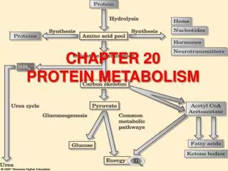
Summary of protein digestion in the human body.Possible fates for amino acid degradation products.
Transamination and Oxidative Deamination: Two steps in degrading amino acids 1) remove -amino group 2) breakdown & process carbon skeleton Release of an amino group is also two steps: 1) Transamination 2) Oxidative deamination
Central role of glutamate: Amino acids: Glutamate, aspartate, alanine & glutamine present in higher concentrations in mammalian cells. Have metabolic functions as well as roles in proteins. Glutamate is the most important, metabolically.
Some transaminases are used for diagnosing disorders: enzyme alanine aminotransferase Escapes in large amounts from dead or dying liver tissue. Measured in blood samples for diagnostic purposes.
Transaminase enzyme aspartate aminotransferase very active enzyme inside heart cells. Also escapes in large amounts from dead or dying heart tissues & enters bloodstream. Measured in blood for diagnosing myocardial infarction.
Trans-deamination (sum it up) Most transaminases share a common substrate and product (oxoglutarate and glutamate) with the enzyme glutamate dehydrogenase. This permits a combined N excretion pathway for individual amino acids: "trans-deamination.” Glutamate has a central role in the overall control of nitrogen metabolism. a.k.a.-ketoglutarate
OxidativeDeamination The glutamate produced from the transamination step is then deaminated by oxidative deamination using the enzyme glutamate dehydrogenase: And away I go! Recycles back to a ketodiacid & releases ammonia
Glutamate dehydrogenase [GluDH] will reversibly convert glutamate to -ketoglutarate and -ketoglutarate to glutamate. Deamination reaction uses NAD+ reverse reaction uses NADPH Uses bothNAD+andNADPH – how to regulate it?
Urea cycle: Ammonium salts (NH4+) are toxic compounds. Oxidative deamination converting glutamate to -ketoglutarate is an easily shifted equilibrium reaction. Ammonium ions building up favors the synthesis of excessive amounts of glutamate, decreasing the Krebs cycle intermediate -ketoglutarate. This in turn decreases ATP production, and that affects the nervous system. The answer is Urea:
The inputs to the urea cycle are NH3, CO2 and aspartic acid and ATP. The outputs are urea, ADP and fumaric acid. The carbonyl group of urea is derived from CO2 Ammonia contributes one of the amine groups on urea
The four-stepurea cycle in which carbamoyl phosphate is converted to urea.
The nitrogen content of the various compounds that participate in the urea cycle. Does it remind you of the Krebs cycle in any way?
Fumarate from the urea cycle enters the Krebs cycle. Aspartate produced from oxaloacetate of the Krebs cycle enters the urea cycle.Oxaloacetate has 4 potential fates: transamination; conversion to glucose; formation of citrate; conversion to pyruvate
Summary: Transamination takes off amine groups from amino acids and forms glutamate (ionized glutamic acid) Amine groups form ammonia when removed in deamination This combines with CO2 & Aspartate. Forms urea, Arginine, & Fumarate
Alternative methods of nitrogen excretion Aquatic species excrete free ammonia through gills. Terrestrial critters produce Urea - very soluble - still needs water for removal via kidneys. Imposes a minimum daily water requirement. Spiders excrete guanine, 5 nitrogen atoms in a small molecule.
Reptiles & birds excrete uric acid – very insoluble purine compound – forms supersaturated solutions. Concentrated urine, supersaturated with uric acid, goes from cloaca into hindgut – uric acid crystalizes & water is reabsorbed. In humans uric acid deposits crystals & causes gout.
Processing Amino Acid Carbon Skeletons Transamination or Oxidative deamination both produce -keto acids Degradation of these carbon skeletons may take several different pathways: Amino acid C skeletons that degrade to form a Krebs cycle intermediate can then be used to make glucose via gluconeogenesis. These are called Glucogenic Amino Acids. Amino acid C skeletons that degrade to form acetyl CoA or Acetoacetyl CoA can form fatty acids or ketone bodies. These are called Ketogenic Amino Acids.
Two are ketogenic: leucine & lysine. Nine are glucogenic. Nine are both because they form pyruvate or have two different degradation products. F ates of C skeletons of 20 amino acids.
Amino Acid Biosynthesis Essential amino acids can be made by plants & bacteria in 7 to 10 steps. We obtain these amino acids by eating plants. 11 Non-essential amino acids synthesized in 1 to 3 steps. Use glycolysis intermediates: 3-phosphoglycerate & pyruvate Krebs cycle intermediates: Oxaloacetate & a-ketoglutarate
Starting materials for biosynthesis of 11 nonessential amino acids: 1 step, 2 steps, or 3 steps Alanine, aspartate, & glutamate use transamination
Phenylalanine & tyrosine degradation: Degradation of phenylalanine starts with conversion to tyrosine catalyzed by phenylalanine hydroxylase. Fumarate & acetoacetate are formed. Fumarate is converted to oxaloacetate for TCA cycle & acetoacetate is converted to acetyl CoA.
Phenylketonuria (PKU): Defective phenylalanine hydroxylase – phenylalanine accumulates in body. Phenylalanine is transaminated to phenylpyruvate. Accumulation of phenylpyruvate leads to severe mental retardation in infants. Persons suffering from phenylketonuria should not consume foods containing high levels of phenylalanine, such as aspartame.
Hemoglobin catabolism Red blood cells contain oxygen carrying pigments of a conjugated protein: Protein part is Globin Non-protein prosthetic group is Heme Heme contains four pyrrole (tetrapyrrole) groups held together by an iron atom Old red blood cells degraded in the spleen. Globin is hydrolyzed into amino acids. Iron atom stored in a protein (ferritin) Tetrapyrrole degraded to bile pigments
Formation of Bile Pigments Ring opens up Uses molecular O2, loses 1 C as CO which fuses to 1% of hemoglobin. Fe3+ grabbed by ferritin to make new hemoglobin Heme Biliverdin
Central methylene bridge of Biliverdin is reduced creating Bilirubin. Bilirubin found to be the major antioxidant in blood
Bilirubin transported to liver. Becomes more water soluble with attachment of glucoronide sugars to propionate side chains. (Glucose + COO- group attached to 6th C) Solubilized bilirubin is excreted in Bile.
Intestinal bacteria help change Bilirubin into Stercobilin and urobilin for excretion.
Changing color of a bruise shows dominant degradation product of heme: either biliverdin or bilirubin Jaundice: when bilirubin accumulates in the blood. Spleen is degrading heme, but liver isn’t removing the products.
Follow each pathway to its various products. All are highly inter-related.
The human body’s response to fasting Remember: the Brain uses Glucose or Ketone bodies for fuel.
The human body’s response to starvation Everything is used to feed the brain Glucose or Ketone bodies.
Review: can you… • Describe the steps in Protein digestion & absorption • Explain how Amino Acids are utilized in the body • Explain Transamination and Oxidative De-amination • Describe The Urea Cycle – purpose and steps • Describe how a.a. Carbon Skeletons are processed • Define and explain Amino Acid Biosynthesis. • Describe the chemical composition of urine. • Explain the relationship and importance of Arginine, • Citrulline, and Nitric Oxide. • Give a detailed description of Hemoglobin Catabolism • Give an overview of the interrelationships among • Carbohydrate, Lipid, and Protein Metabolic Pathways.