Download
1 / 66
660 likes | 842 Vues
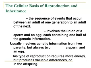
Chapter 8: The Cellular Basis of Reproduction and Inheritance. Introduction Stages of an Organism’s Life Cycle: Development : All changes that occur from a fertilized egg or an initial cell to an adult organism.

E N D
Chapter 8: The Cellular Basis of Reproduction and Inheritance
Introduction Stages of an Organism’s Life Cycle: Development: All changes that occur from a fertilized egg or an initial cell to an adult organism. Reproduction: Production of offspring that carry genetic information in the form of DNA, from their parents. Two types of reproduction: 1. Sexual Reproduction 2. Asexual Reproduction
Types of reproduction: 1. Sexual Reproduction: • Most common type of animal reproduction. • Male and female gametes or sex cells (sperm and egg cell) join together to create a fertilized egg or zygote. • The offspring has genetic information from both parents. • Offspring are genetically different from each parents and their siblings. • Advantages: • Ensures genetic diversity of offspring. • Population more likely to survive changing environment. • Disadvantages: • Cannot reproduce without a partner of opposite sex. • Considerable time, energy, and resources spent to find a suitable mate. • Parents only pass on 1/2 (50%) of their genetic information to each offspring.
2. Asexual Reproduction: • Production of offspring by a single parent through: • Splitting: Binary fission in bacteria. • Budding: Yeasts, plants • Fragmentation: Sea stars • Parthenogenesis: “Virgin birth”. Several insect species. • Offspring inherit DNA form one parent only. • Offspring are genetically identical to parent and siblings, unless mutations occur. • Advantages: • Can reproduce without a partner of opposite sex. • Don’t spend time, energy, and resources to find a suitable mate. • Parents pass on 100% of their genetic information to each offspring. • Disadvantage: • No genetic diversity of offspring. • Population less likely to survive changing environment.
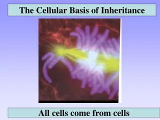
Cells Only Arise from Preexisting Cells New cells are made through cell division: • Unicellular organisms (Bacteria, protozoa): Division of one cell into two new organisms through binary fission or mitosis. • Multicellular organisms (Plants, animals): If sexual reproduction: 1.Growth and development from zygote or fertilized egg. • Original cell divides by mitosis to produce many cells, that are genetically identical to first cell. Cells later develop specific functions (differentiation). 2.Reproduction requires: • Meiosis: Special type of cell division that will generate gametes or sex cells, with 50% of individual’s genetic material.
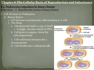
Bacteria (Procarytoes) Reproduce Asexually by Binary Fission Features of Bacterial DNA • Single, relatively small circular chromosome: About 3-5 million nucleotide base pairs • Contains only about 5-10,000 genes • Binary fission • Single circular DNA is replicated • Bacterium grows to twice normal size • Cell divides into two daughter cells • Each daughter cell with an identical copy of DNA • Rapid process, as little as 20 minutes.
Eucaryotic cell division is a more complex and time consuming process than binary fission Features of Eucaryotic DNA 1. DNA is in multiple linear chromosomes. • Unique number for each species: • Humans have 46 chromosomes. • Cabbage has 20, mosquito 6, and fern over 1000. 2. Large Genome: Up to 3 billion base pairs (humans) • Contains up to 50,000-150,000 genes • Human genome project is determining the sequence of entire human DNA. 3. DNA is enclosed by nuclear membrane. Correct distribution of multiple chromosomes in each daughter cell requires a much more elaborate process than binary fission.
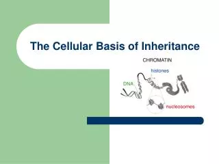
DNA: Found as Chromosomes or Chromatin Chromosomes Chromatin Tightly packaged DNA Unwound DNA Found only during cell Found throughout cell divisioncycle DNA is not being used DNA is being used for macromolecule for macromolecule synthesis. synthesis.
Sequence of events from the time a cell is formed, until the cell divides once again. • Before cell division, the cell must: • Precisely copy genetic material (DNA) • Roughly double its cytoplasm • Synthesize organelles, membranes, proteins, and other molecules. • Cell cycle is divided into two main phases: • Interphase: Stage between cell divisions • Mitotic Phase: Stage when cell is dividing Cell Cycle of Eucaryotic Cells
The Life Cycle of a Eucaryotic Cell: Interphase: Time between cell divisions. • Most cells spend about 90% of their time in interphase. • Cells actively synthesize materials they need to grow. • Chromosomes are duplicated. • Interphase can be divided into three stages: 1. G1 phase:Just after cell division. Cell grows in size, increases number of organelles, and makes proteins needed for DNA synthesis. 2. S phase: DNA replication. Single chromosomes are duplicated so they contain two sister chromatids. 3. G2 phase: Just before cell division. Protein synthesis increases in preparation for cell division.
Duplication of Chromosomes During S stage of Interphase DNA replication during S stage of Interphase Single chromosome Two identical sister chromatids joined by a centromere ( )
The Life Cycle of a Eucaryotic Cell: Mitosis:The process of eucaryotic cell division. • Most cells spend less than 10% of time in mitosis. • Mitosis is divided into four stages: 1. Prophase: Cell prepares for division. 2. Metaphase: Chromosomes line up in “middle” of cell. 3. Anaphase: Sister chromatids split and migrate to opposite sides of the cell. 4. Telophase: DNA is equally divided into two new daughter cells. Cytokinesis usually occurs. • Cytokinesis: Division of cytoplasm. Mitotic Phase: Mitosis + Cytokinesis
Mitosis: The Stages of Cell Division 1. Prophase • Chromatin condenses into chromosomes, which appear as two sister chromatids joined by a centromere. • Nucleoli disappear. • Nuclear envelope breaks apart. • In animal cells, mitotic spindle begins to form as mictotubules grow out of two centrosomes or microtubuleorganizingcenters (MTOCs). • Each centrosome is made up of a pair of centrioles. • Microtubules attach to kinetochores on chromatids and begin to move chromosomes towards center of cell. • Centrosomes begin migrating to opposite poles of cell.
Mitosis: The Stages of Cell Division 2. Metaphase • Short period in which chromosomes line up along equatorial plane of cell (metaphase plate). • Chromosomes are completely condensed and easy to visualize. • Mitotic spindle is fully formed. • Kinetochores of sister chromatids face opposite sides and are attached to spindle microtubules at opposite ends of the cell.
Metaphase, Anaphase, and Telophase of Mitosis in an Animal Cell
Mitosis: The Stages of Cell Division 3.Anaphase • Centromeres of sister chromatids begin to separate. • Each chromatid is now an independent daughter chromosome. • The separate chromosomes are pulled toward opposite ends by spindle microtubules, attached to the kinetochores. • Cell elongates as poles move farther apart. • Anaphase ends when a complete set of chromosomes reaches each pole.
Mitosis: The Stages of Cell Division 4. Telophase • Cell continues to elongate. • Cell returns to interphase conditions: • A nuclear envelope forms around each set of chromosomes. • Chromosomes uncoil, becoming chromatin threads. • Nucleoli reappear. • Spindle microtubules disappear. • Cytokinesis usually occurs at the end of this stage
Mitotic Phase: Mitosis + Cytokinesis • Cytokinesis • The division of cytoplasm to produce two daughter cells. Usually begins during telophase. • In animal cells: Division is accomplished by a cleavage furrow that encircles the cell like a ring in the equator region. • In plant cells: Division is accomplished by the formation of a cell plate between the daughter cells. Each cell produces a plasma membrane and a cell wall on its side of the plate.
Cytokinesis in Animal and Plant Cells Animal Cell Plant Cell
External Factors Control Mitosis 1. Anchorage • Most cells cannot divide unless they are attached to a solid surface. • May prevent inappropriate growth of detached cells 2. Nutrients and growth factors • Lack of nutrients can limit mitosis • Growth factors: Proteins that stimulate cell division. 3. Cell density • Density-dependent inhibition: Cultured cells will stop dividing after a single layer covers the petri dish. Mitosis is inhibited by high cell density. • Cancer cells do not demonstrate density inhibition
Density Dependent Inhibition of Mitosis Normal Cells Stop Dividing at High Cell Density Cancer Cells are Not Inhibited by High Cell Density
There are three critical points at which the cell cycle is controlled*: 1. G1 Checkpoint: Prevents cell from entering S phase and duplicating DNA. • Most important checkpoint. • Amitotic cells (muscle and nerve cells) are frozen here. 2. G2 Checkpoint: Prevents cell from entering mitosis. 3. M Checkpoint: Prevents cell from entering cytokinesis. *Cells must have proper growth factors to get through each checkpoint. Cell-Cycle Control System
Cell Division is Controlled at Three Key Stages Growth factors are required to pass each checkpoint
Cancer kills 1 in 5 people in the United States. • Cancer cells divide excessively and invade other body tissues. • Tumor: Abnormal mass of cells that originates from uncontrolled mitosis of a single cell. • Benign tumor:Cancer cells remain in original site. Can easily be removed or treated • Malignant tumor:Cancer cells have ability to “detach” from tumor and spread to other organs or tissues • Metastasis:Spread of cancer cells form site of origin to another organ or tissue. • Tumor cells travel through blood vessels or lymph nodes. Cancer is a Disease of the Cell Cycle
Functions of Mitosis in Eucaryotes: 1.Growth:All somatic cells that originate after a new individual is created are made by mitosis. 2. Cell replacement:Cells that are damaged or destroyed due to disease or injury are replaced through mitosis. 3. Asexual Reproduction:Mitosis is used by organisms that reproduce asexually to make offspring.
Chromosomes are matched in homologous pairs Homologous Chromosomes: • Eucaryotic chromosomes come in pairs. Normal humans have 46 chromosomes in 23 pairs. • One chromosome of each pair comes from an individual’s mother, the other comes from the father. • Homologous chromosomes carry genes that control the same characteristics. • Examples: Eye color, blood type, flower color, or height • Locus: Physical site on a chromosomes where a given gene is located. • Allele: Different forms of the same gene. • Example: Alleles for blood types A, B, or O.
Homologous Pair of Chromosomes: One Comes From Each Parent
Homologous Chromosomes: Code for the Same Genetic Traits, but Have Different Alleles
There are two types of chromosomes: 1. Autosomes: Found in both males and females. • In humans there are 22 pairs of autosomes. • Autosomes are of the same size and are homologous. 2. Sex Chromosomes:Determine an individual’s gender. • One pair of chromosomes (X and Y). • The X and Y chromosomes are not homologous. • The X chromosome is much larger than the Y chromosome and contains many genes. • The Y chromosome has a small number of genes. • In Humans and other mammals females are XX and males are XY.
Chromosomes of Normal Human Male:44 (22 Pairs) Autosomes + XY
Normal Genetic Complement of Humans: Females: 44 autosomes (22 pairs) + XX Males: 44 autosomes (22 pairs) + XY Note: In most cases, having additional or missing chromosomes is usually fatal or causes serious defects. Down’s syndrome: Trisomy 21. Individual’s with an extra chromosome 21. Most common chromosomal defect (1 in 700 births in U.S.). Mental retardation, mongoloid facial features, heart defects, etc.
Gametes have a single set of chromosomes Humans have two sets of chromosomes, one inherited from each parent. • Diploid Cells: Cells whose nuclei contain two homologous sets of chromosomes (2n). • Somatic cells are diploid (almost all cells in our body). • In humans the diploid number (2n) is 46. • Haploid Cells:Cells whose nuclei contain a single set of chromosomes (n). • Gametes are haploid (egg and sperm cells). • In humans the haploid number (n) is 23. Fertilization: Haploid egg fuses with a haploid sperm to form a diploid zygote (fertilized egg).
Meiosis Produces Haploid Gametes From Diploid Parents Fertilization Produces Diploid Offspring from Haploid Gametes
Mitosis versus Meiosis MitosisMeiosis Onecell divisionTwosuccessive cell divisions Produces two(2) cellsProducesfour (4) cells Produces diploidcellsProduceshaploidgametes Daughter cells are geneticallyCells are geneticallydifferentfrom identicalto mother cell mother cell and each other No crossing over Crossing over* Functions: Growth, Functions: Sexual reproduction cell replacement, and asexual reproduction *Crossing over: Exchange of DNA between homologous chromosomes.
Meiosis: Generates haploid gametes • Reduces the number of chromosomes by half, producing haploid cells from diploid cells. • Also produces genetic variability, each gamete is different, ensuring that two offspring from the same parents are never identical. • Two divisions: Meiosis I and meiosis II. Chromosomes are duplicated in interphase prior to Meiosis I. • Meiosis I:Separates the members of each homologous pair of chromosomes. Reductive division. • Meiosis II: Separates chromatids into individual chromosomes.
STAGES OF MEIOSIS Interphase: Chromosomes replicate Meiosis I: Reductive division. Homologous chromosomes separate Meiosis II: Sister chromatids separate
Meiosis I:Separation of Homologous Chromosomes 1. Prophase I: • Most complex phase of meiosis (90% of time) • Chromatin condenses into chromosomes. • Nuclear membrane and nucleoli disappear. • Centrosomes move to opposite poles of cell and microtubules attach to chromatids. • Synapsis: Homologous chromosomes pair up and form atetradof 4 sister chromatids. • Crossing over:DNA is exchanged between homologous chromosomes, resulting ingenetic recombination. Unique to meiosis. • Chiasmata: Sites of DNA exchange.
Meiosis I:Separation of Homologous Chromosomes 2. Metaphase I: • Chromosome tetrads (homologous chromosomes) line up in the middle of the cell. • Each homologous chromosome faces opposite poles of the cell.
Stages of Meiosis: Meiosis I 3. Anaphase I: • Chromosome tetrads split up. • Homologous chromosomesof each pairseparate, moving towards opposite poles. • Random assortment: One chromosome from each homologous pair is shuffled into the two daughter cells, randomly and independently of the other pairs. • Random assortmentincreases genetic diversityof offspring. Possible combinations:2n. • One human cell can generate 223 or over 8.3 million different gametes by random assortment alone.