
How is apoptotic cell recognized and engulfed?
How is apoptotic cell recognized and engulfed?. Wild-type. ced-1. In 1983, E. Hedgecock isolated two cell death mutants ( ced-1 and ced-2 ) which are pivotal for identification of the other cell death mutants. Wild-type. ced-x?.
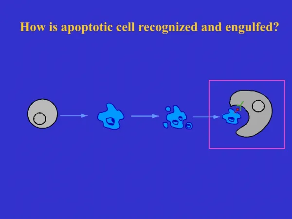

How is apoptotic cell recognized and engulfed?
E N D
Presentation Transcript
Wild-type ced-1 In 1983, E. Hedgecock isolated two cell death mutants (ced-1 and ced-2) which are pivotal for identification of the other cell death mutants.
Wild-type ced-x? Similar screens to look for more engulfment defective mutants Did not identify any additional mutants except more ced-1 mutations Why?
Why no additional mutants were found? • Only one gene is needed • B) Phenotypes are too weak and thus mutations are difficult to isolate • C) The graduate students did not try hard enough • D) Maternal effect • E) Other engulfment mutants are all lethal.
Rare case P0 m/+ x m/+ m/+ x m/+ F1 m/m m/m mutant Phenotype wild-type F2 m/m m/m mutant mutant Phenotype Maternal Effects A maternal effect is the phenomenon where the genotype of a mother is expressed in the phenotype of its offspring, unaltered by paternal genetic influence
Maternal Effects P0 ced-1/+ ced-2/+ F1 ced-1/ced-1 ced-2/ced-2 Many corpses wild-type Phenotype F2 Many corpses Many corpses Are there more engulfment genes that have maternal effects?
EMS P0 tra-2(n1106) tra-2(n1106); m/+ rare F1 F2 +/+ m/+ Egl suppressor m/m m/+ Regular Genetic Screens Look for rare F2 mutants
Maternal Effect Screens EMS P0 tra-2 ( n1106 ) ; m/+ Rare tra-2 n1106 ( ) F1 F2 +/+ m/+ No suppressor m/m m/+ F3 suppressor R. Ellis and R.H. Horvitz performed F3 screen and identified mutations in four additional genes affecting cell corpse engulfment ced-5, ced-6, ced-7 and ced-10
Cell migration Two partially redundant pathways control cell corpse engulfment in C. elegans ced-1 ced-6 ced-7 cell corpse engulfment ced-2 ced-5 ced-10 ced-12
Why are ced-2, -5, -10, -12 mutants defective in both corpse engulfment and cell migration? • It is purely coincidental. • These genes regulate a cellular process common to both corpse engulfment and cell migration • Cell migration is a required step for corpse engulfment • Corpse engulfment is a required step for cell migration • None of above
Cell corpse engulfment Cell-cell signaling between the dying cell and the phagocytic cell
Cell corpse engulfment Cell-cell signaling between the dying cell and the phagocytic cell
ced-2, 5, 10, 12 encode proteins regulate cytoskeleton reorganization ced-2 CRK II (SH2, SH3) ced-5 DOCK180 ced-10 Rac I (GTPase) ced-12 regulator of Rac
CED-7 ? CED-2 CED-5 CED-12 CED-10 Apoptotic cell cytoskeleton Engulfing cell
Cell migration Two partially redundant pathways control cell corpse engulfment in C. elegans ced-1 ced-6 ced-7 cell corpse engulfment ced-2 ced-5 ced-10 ced-12
CED-1 is a membrane protein CED-1 may recognize apoptotic cells CED-1 is internalized by engulfing cells CED-1 cluster on the surface of apoptotic cells All of the above CED-1::GFP ced-1 EGF-like receptor CED-1::GFP
ced-1 and ced-7 might be involved in cell corpse recognition ced-1 EGF-like receptor ced-7 ABCtransportor ced-6 phosphotyrosine-binding protein CED-1::GFP CED-1::GFP In ced-7(lf) mutants, CED-1::GFP no longer encloses apoptotic cells
ced-7 may be involved in recognizing or exposing an “eat-me” signal ced-1 may be involved in recognizing an “eat-me” signal ced-6 phosphotyrosine-binding protein
CED-6 cytoskeleton CED-7 CED-1 CED-7 ? CED-2 CED-5 CED-12 CED-10 Apoptotic cell cytoskeleton Engulfing cell What are the “eat-me” signals?
Normal cell Phosphatidylserine(PS) Apoptosis Dying cell Engulfment PS is restricted to the inner leaflet of plasma membrane and its externalization can trigger phagocytosis Phagocyte
* * * * * * * * * * Secreted AnxV::GFP 23
CED-8, an unexpected activator of apoptotic PS externalization • ced-8 was first identified as a gene involved in regulating the timing of apoptosis and encodes a homologue of human XK transporters (Stanfield and Horvitz, Mol Cell 2000) Out Out CED-8 acCED-8 In In CED-3 CED-3 cleavage Living cells Dying cells
Ectopic expression of acCED-8 is sufficient to induce PS externalization in all living cells in C. elegans Surface-exposed PS is labeled by the PS-binding Lactadherin::GFP Chen et al., Nature Communications 2013.
Clearance of cell corpses is significantly compromised in the ced-8(-/-) mutant
Death stimulus PS PS WAH-1 promotes nuclear and cell surface apoptotic events through CPS-6 and SCRM-1 SCRM-1 WAH-1 CPS-6
cps-7 cps-9 cps-12 cps-13 cps-14 cytoskeleton Multiple cps genes mediate recognition and engulfment of apoptotic cell corpse PS (phosphatidylserine)
PSR-1 is a PS binding protein Yang et al., (2015) Nature Communications
cps-7 cps-9 cps-12 psr-1 cps-13 cps-14 PSR-1 may transduce the “PS eat-me” signal through CED-5 and CED-12 signaling pathway CED-5 PS CED-10 CED-2 CED-12 Wang et al., (2003) Science 302: 1563-1566
How do living cells maintain PS asymmetry? What happens if PS asymmetry is disrupted in living cells?
Aminophospholipid translocases are implicated in restricting PS to the inner leaflet of plasma membrane There are 6 aminophospholipid translocases in C. elegans, which are annotated as tat genes (transbilayer amphipath transporter)
Genetic inactivation of tat-1 but not other tat genes causes stronger PS exposure on the surface of living cells DIC Annexin V Hoechst DIC Annexin V Hoechst tat-1(-/-) tat-4(-/-) tat-2(-/-) tat-5(-/-) tat-3(-/-) tat-6(-/-) tat-1 functions to restrict PS to the inner leaflet of plasma membrane Darland-Ransom et al. Science 320, 528, 2008
Can externalized PS in living cells induce phagocytosis? AVM PLM ALM PHA/PHB PVM HSN ADE VC Pida-1GFP Pmec-4GFP % animals missing at least one touch cell % animals missing at least one neurons WT 1% WT1% tat-1(lf) 19% tat-1(lf) 24% tat-3(lf) 1% tat-3(lf) 2% Inactivation of tat-1 causes random loss of living cells
CED-6 cytoskeleton CED-7 CED-1 CED-7 ? CED-2 CED-5 CED-12 CED-10 Apoptotic cell cytoskeleton Engulfing cell Two partially redundant pathways promote removal of apoptotic cells in C. elegans ced-6 Exposed PS ced-1 ced-7 PSR-1 PS ced-2 Exposed PS ced-10 ced-5 psr-1 ced-12
Living cells in the tat-1 mutant are removed by a phagocytic mechanism AVM PLM ALM PHA/PHB PVM HSN ADE VC Pmec-4GFP Pida-1GFP % animals missing at least one touch cell % animals missing at least one neurons WT1% WT 1% tat-1(lf) 19% tat-1(lf) 24% ced-1(lf) 2% ced-1(lf) 0% ced-1(lf); tat-1(lf) 1% ced-1(lf); tat-1(lf) 0% psr-1(lf) 2% psr-1(lf) 1% tat-1(lf);psr-1(lf) 2% tat-1(lf);psr-1(lf) 2%


