Lecture 18 The Urinary System
210 likes | 672 Vues
Lecture 18 The Urinary System. 5 Functions of the Urinary System. Regulate blood volume and blood pressure: by adjusting volume of water lost in urine releasing erythropoietin and renin Regulate plasma ion concentrations:

Lecture 18 The Urinary System
E N D
Presentation Transcript
5 Functions of the Urinary System • Regulate blood volume and blood pressure: • by adjusting volume of water lost in urine • releasing erythropoietin and renin • Regulate plasma ion concentrations: • sodium, potassium, and chloride ions (by controlling quantities lost in urine) • calcium ion levels (through synthesis of calcitriol) • Help stabilize blood pH: • by controlling loss of hydrogen ions and bicarbonate ions in urine • Conserve valuable nutrients: • by preventing excretion while excreting organic waste products • Assist liver to detoxify poisons
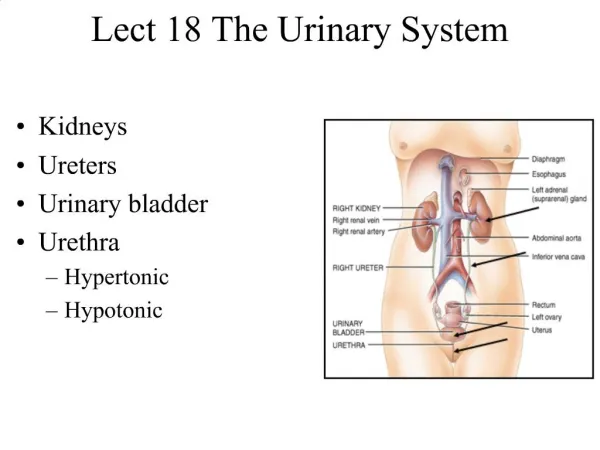
Urinary System Organs • Kidney – produces urine • Urinary bladder – provides a temporary storage reservoir for urine • Paired ureters – transport urine from the kidneys to the bladder • Urethra – transports urine from the bladder out of the body
Quick Facts on the Kidneys • The kidneys filter 200 liters of blood daily, allowing toxins, metabolic wastes, and excess ions to leave the body in urine • Approximately one-fourth (1200 ml) of systemic cardiac output flows through the kidneys each minute • All the blood in the body is filtered 60 times everyday (2½ times every hour)
Internal Anatomy of the Kidney • A frontal section shows three distinct regions • Cortex – the light colored, granular outer region • Medulla – the inner region that exhibits cone-shaped medullary (renal) pyramids • 6-18 Pyramids are made up of parallel bundles of urine-collecting tubules • Renal columns are inward extensions of cortical tissue that separate the pyramids • A Lobe is a medullary pyramid and its surrounding capsule • Papillae – drain urine from a lobe into a minor calyx • Draining passages • Minor Calyces – small branches between the lobes and the major calyces • Major calyces – large branches of the renal pelvis • Renal pelvis – flat, funnel-shaped tube lateral to the hilus within the renal sinus • Urine flows through the pelvis and ureters to the bladder
Renal Pyramids Contain Nephrons A nephron is composed of three regions • A Filter (the Renal Corpuscle): • Glomerular capsule encloses a • Glomerulus a fine network of capillaries • A Tubule (3 sections): • Proximal Convoluted Tubule (PCT) • Loop of Henle is bent back on itself in the center • Distal Convoluted Tubule (DCT) • Site of reabsorption & secretion • A Collecting Duct • Water conservation device
Filtration Membrane • The glomerulus is a filter that lies between the blood and the interior of the glomerular capsule • It is composed of three layers • Fenestrated endothelium of the glomerular capillaries • Visceral membrane of the glomerular capsule (podocytes) • Basement membrane composed of fused basal laminae of the other layers Figure 25.7a
2 Types of Nephrons • Cortical nephrons – 85% of nephrons; located in the cortex • Juxtamedullary nephrons: • Are located at the cortex-medulla junction • Have loops of Henle that deeply invade the medulla • Have extensive thin segments • Are involved in the production of concentrated urine
Capillary Beds • Blood pressure in the glomerulus is high because: • Arterioles are high-resistance vessels • Afferent arterioles have larger diameters than efferent arterioles • Fluids and solutes are forced out of the blood throughout the entire length of the glomerulus • Peritubular beds are low-pressure, porous capillaries adapted for absorption that: • Arise from efferent arterioles • Cling to adjacent renal tubules • Empty into the renal venous system • Vasa recta – long, straight efferent arterioles of juxtamedullary nephrons • Every nephron has two capillary beds • Glomerular capillaries • Peritubular capillaries • Each glomerulus is: • Fed by an afferent arteriole • Drained by an efferent arteriole
Juxtaglomerular Apparatus (JGA) • The distal tubule lies against the afferent (sometimes efferent) arteriole • Arteriole walls have juxtaglomerular (JG) cells • Enlarged, smooth muscle cells • Have secretory granules containing renin • Act as mechanoreceptors • Macula densa cells • Tall, closely packed distal tubule cells • Lie adjacent to JG cells • Function as chemoreceptors or osmoreceptors
Mechanisms of Urine Formation • The kidneys filter the body’s entire plasma volume 60 times each day • The filtrate: • Contains all plasma components except protein • Loses water, nutrients, and essential ions to become urine • The urine contains metabolic wastes and unneeded substances
Mechanisms of Urine Formation • Urine formation and adjustment of blood composition involves three major processes • Glomerular filtration • Tubular reabsorption • Tubular secretion
Glomerular Filtration Rate (GFR) • The total amount of filtrate formed per minute by the kidneys • Factors governing filtration rate at the capillary bed are: • Total surface area available for filtration • Filtration membrane permeability • Net filtration pressure (NFP) • Changes in GFR normally result from changes in glomerular blood pressure • GFR is directly proportional to the NFP
Glomerular Filtration • The glomerulus is more efficient than other capillary beds because: • Its filtration membrane is significantly more permeable • Glomerular blood pressure is higher • It has a higher net filtration pressure • Plasma proteins are not filtered and are used to maintain osmotic pressure of the blood • If the GFR is too high: • Needed substances cannot be reabsorbed quickly enough and are lost in the urine • If the GFR is too low: • Everything is reabsorbed, including wastes that are normally disposed of • Three mechanisms control the GFR • Renal autoregulation (intrinsic system) • Neural controls • Hormonal mechanism (the renin-angiotensin system)
Absorption in Renal Tubules and Collecting Ducts • PCT reabsorbs substances including: • Sodium, all nutrients, cations, anions, and water • Urea and lipid-soluble solutes • Small proteins • Loop of Henle reabsorbs: • H2O, Na+, Cl, K+ in the descending limb • Ca2+, Mg2+, and Na+ in the ascending limb • DCT absorbs: • Ca2+, Na+, H+, K+, and water • HCO3 and Cl • Collecting duct absorbs: • Water and urea
(Filtrate ~ 100 mOsm) Active reabsorption Na+ Na+/K+-Cl- symporter Permeable to Water Not to Solutes ~ 300 mOsm Permeable to Solutes Not to Water Na+-H- antiporter Additional 10% of original water passively reabsorbed Additional 40% of original NaCl reabsorbed Passive reabsorption Na+ ~ 1200 mOsm 200 mOsm Loop of Henle How Kidney Tubules Create an Osmotic Gradient and produce the initial dilute urine Proximal Convoluted Tubule
Vasa Recta: Countercurrent Exchange • The vasa recta is a countercurrent exchanger that: • Delivers blood to the cells in the area • While maintaining the osmotic gradient
IF plasma > 300 mOsm Variable Process Producing concentrated urine Permeable to Water Not to Solutes Distal Convoluted Tubule Additional 10% of original water passively reabsorbed Medullary Collecting Duct Water Reabsorption Pituitary releases ADH Water (& urea) Reabsorbed Insertion of aquaporins into luminal membrane 200 mOsm How Kidney Tubules Produce Concentrated Urine Common Process Producing initial dilute urine ~ 300 mOsm (Filtrate ~ 100 mOsm) Proximal Convoluted Tubule Permeable to Solutes Not to Water Additional 40% of original NaCl reabsorbed ~ 1200 mOsm Loop of Henle
Summary of Urine Concentration • Two things are required to concentrate urine • A high osmotic gradient in the kidney’s medulla • The presence of ADH • The loop of Henle is the primary structure responsible for maintaining the hypertonic medullary interstitium • The presence of ADH is mediated by blood plasma osmolality as sensed by the hypothalamus • The final concentration of urine cannot exceed the mOsm level of the most concentrated part of the hypertonic medullary interstitium.
Summary Quiz on Concentration of Urine 6. ~ 300 mOsm When the DCT in this region is permeable to water, will water flow into or out of the tubule? Distal Convoluted Tubule Proximal Convoluted Tubule (Filtrate ~ 100 mOsm) 2. 7. What flows out of the filtrate in the ascending limb? What hormone mediates water permeability in the tubules? Medullary Collecting Duct 1. What flows out of the filtrate in the descending limb? 8. 3. What condition stimulates its release? What hormone mediates the process in the previous question? 9. In addition to water, what solute flows out of the duct in this region? 4. Does the filtrate have higher osmolality in the Descending or Ascending limb? ~ 1200 mOsm 10. What does this solute help maintain? Loop of Henle 5. This difference in osmolality is called the? 11. As water is removed from the filtrate what happens to the filtrate volume?