Download
1 / 50
500 likes | 513 Vues
International Conference Diagnosis & Treatment of Inner Ear Disorders Genetics of deafness. Lech Korniszewski The Medical University of Warsaw Institute of Physiology and Pathology of Hearing. Hearing loss – incidence:.

E N D
International Conference Diagnosis & Treatment of Inner Ear DisordersGenetics of deafness Lech Korniszewski The Medical University of Warsaw Institute of Physiology and Pathology of Hearing
Hearing loss – incidence: 6-8% of population –when all causes are combined hearing loss – most common birth defect 1 in 1000 newborns are deaf 1 in 300 children are affected with congenital hearing loss of a lesser degree additional 1 in 1000 become profoundly hearing impaired before adulthood
Genetic hearing loss approximately 1% of all human genes are involved in the hearing process inheritance: autosomal recessive autosomal dominant X-linked mitochondrial • allelic mutatione in some genes can cause recessive and dominant hearing loss • mutations in the same gene may cause syndromic or nonsyndromic hearing loss • recessive hearing loss may be caused by a combination of two mutations in differrent genes from the same functional group
Syndromic hearing loss Over 400 syndromes have been described in which hearing lossis a component part. There are many factors that make specificsyndrome diagnosis difficult: • The rarity of most of these syndromes (lack personal experience) • Variability of clinical expression • Genetic heterogeneity (a single phenotype may be result of different genes mutations) • Pleiotropy (single gene may cause many different phenotypic effects)
Waardenburg syndromes • Bilateral or unilateral sensorineural hearing loss in association with defects in tissues derived from neural crest cells • pigmentary abnormalities hair, skin and eyes • hearing loss is due to defective migration of melanocytes info the intermediate layer of the stria vascularis • genetically heterogeneous; inheritance AD • four clinical subtypes
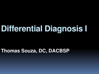
Transcription factor hierarchy in Waardenburg syndrome: regulation of MITF expression by SOX10 and PAX3 EDN3 EDNRB3 WS 1 WS 3 PAX 3 SOX 10 WS 4 transactivation WS 2 MITF transactivation melanocyte tyrosinase
Branchio-oto-renal syndrome Hearing loss conductive, sensorineural or mixed; Branchial cysts and fistulae, external ear malformations, renal dysplasia or hypoplasia. Some patients also eye anomalies Gene EYA1 on 8q13.3; encoded molecule – transcription factor. Inheritance autosomal dominant. Genetically heterogenous (second BOR locus on 1p31)
Treacher-Collins syndrome Hearing loss conductive, sensorineural or mixed; Clinical features: down-slanting palpebral fissures, malformation of external and middle ears, sparse lower eyelashes and colobomata of lower eyelids, malar hypoplasia. Gene TCOF; encoded nuclear cytoplasmic transport protein Inheritance autosomal dominant
Usher syndromes • Syndromic association of hearing loss with retinitis pigmentosa • Accounts 2-4% of all cases of profound deafness and 50% of the deaf-blind population • Inheritance autosomal recessive. Genetic heterogeneity high – more than 12 loci Clinically three main types:
Pendred syndrome Sensorineural deafness, goiter and malformation of the inner ear • Hearing loss is most frequently profound, variable in its onset, rapidly progressive • Goiter results from a specific defect in the organification of iodine (abnormal release of iodine trapped by thyroid after administration of perchlorate) • Malformation of the inner ear in 86% of cases: dilatation of the vestibular aqueduct and endolymphatic sacs, Mondini malformation Inheritance autosomal recessive Mutation of SLC26A4 gene encoding pendrin – protein primarily involved in transport of chloride and iodide ions. Nonsyndromic deafness DFNB4 also result from mutation in the SLC26A4 gene.
Jervell and Lange-Nielsen syndrome • Congenital sensorineural hearing loss and prolongation of the QT interval on electrocardiogram • Hearing loss initially involves the high frequencies and progress to become a profound • Prolongation of QT reflect a defect in cardiacrepolarization. This can lead to recurrent attacks of syncope, ventricular arrhythmia and possible sudden death. • Mutation in genes KCNQ4, KCNE1 coding potassium chanels (K+ active transport in outer hair cells) • Inheritance autosomal recessive
Alport syndrome • Association of sensorineural high frequency hearing loss with progressive nephritis. Anterior lenticonus, macular flecks, cataracts • Gene mutation: COL4A5, COL4A3, COL4A4 coding tissue specific polypeptide subunits of collagen • The subunits are expressed in the basilar membrane, spiral ligament and basement membranes of the stria vascularis • Genetically heterogeneous. Inheritance X-linked dominant and autosomal recessive
Stickler syndrome • sensorineural hearing loss, high frequency, progressive • Myopia, retinal detachment • Arthropathy • Mid-face hypoplasia, cleft palate, micrognathia • Gene defect: COL2A1, COL11A1, COL11A2 • Inheritance autosomal dominant
Hearing loss caused by mutation in GJB2(connexin deafness) • most common cause of hearing loss in many populations • deafness usually stable, onset is nearly always prelingual (but not necessarily congenital); hearing may be normal at birth and hearing loss progress rapidly during first few month of life (some babies may pass neonatal hearing screening but become deaf during infancy) • GJB2 encodes a gap junction protein – connexin 26 • most common mutation is a deletion of single guanine – 35delG (70% mutant alleles, carrier frequency 2-3%) • mutation 35delG in thought rather a founder effect not hot-spot deletion • GJB2 mutations may also be a rare cause of autosomal dominant deafness – syndromic and nonsyndromic (DFNA3). Specific mutation: - hyperkeratosis palmoplantaris - mutilating keratoderma – (Vohwinkel sy.) - keratoderma – ichthyosis – deafness (KID sy.)
Screening GJB2 should be offering as part of the routine work-up in the diagnosis of all cases of non-syndromic deafness of unknown cause. Rationale: - common cause of hearing impairment - phenotype unremarkable and variable - small coding region - common mutations in some populations - enables accurate genetic information to begiven to families disadvantages: counselling difficult with missense and heterozygous mutation
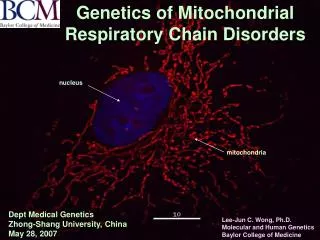
Mitochondrial hearing loss • Sensorineural hearing loss is present in 40-70% patients with mitochondrial disorders and canbe syndromic or non-syndromic. • Mitochondrial mutations are transmitted exclusively through the maternal line and demonstrate complete (or nearly complete) homoplasmy. • Up to 20% patients receiving aminoglycosides experience hearing impairment. 50% of those carry the 12S ribosomal RNA mutation 1555A>G. • Mitochondrial hearing loss may be syndromic: Kearns-Sayre sy., MELAS, maternally inherited diabetes and deafness, and others • Pathogenesis of mitochondrial hearing loss is based on high ATP requirement in the cochlear hair cells. A reduction of available ATP caused by dysfunction of the mitochondrial oxidative phosphorylation results in disturbances of the ionic gradient in the inner ear.