Download
1 / 30
310 likes | 433 Vues
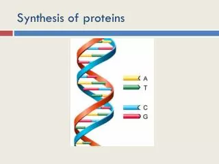
The genetic code, the molecular mechanism of translation and the synthesis of proteins. The information encoded in DNA is transcribed into RNA and finally translated into the sequence of proteins.

E N D
The genetic code, the molecular mechanism of translation and the synthesis of proteins The information encoded in DNA is transcribed into RNA and finally translated into the sequence of proteins. The genetic unit coding for one single amino acid is a codon. One gene codes for one proteins, one cistron for one polypeptide chain. As many proteins consist of only one polypeptide chain, many genes have only one cistron. Decoding of the information present in mRNA is carried out by ribosomes (rs). Amino acids used for the polypeptide synthesis are transported to the ribosomes by tRNA molecules (charged tRNAs). Aminoacylation of tRNAs is catalyzed by very specific aminoacyl-tRNA synthases. The energy required for protein synthesis is supplied by macroerg ATP and GTP molecules.
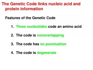
The genetic code coding for 20 amino acids (+ start and stop) 4 nucleotides are not enough using 2 letters the number of combinations is still too few (16) 3 letter combinations are plentiful: 64. How many are used? Does the code have commas or isthecodecontinuous? (ATGcGTCaGGTaTTC...)? Is the code overlapping or not? (ATGTCACAA)? GTTAGC Experiments with intercalating mutagens suggested a 3 letter code: activity of a bacterial enzyme was lost after mutagen treatment a second mutation in the same gene did not change the situation some of the further mutagenized bacteria recovered (part of) the enzyme activity
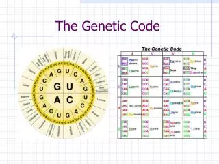
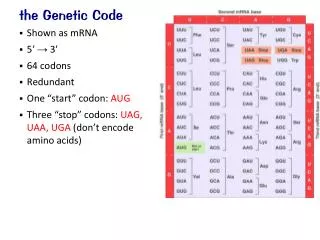
Deciphering the code Artificial mRNAs (synthetic oligo-ribonucleotides) conduct the synthesis of peptides: Poly rU: phenylalanin, poly rC: prolin, poly rA: lysine, poly rG: no product (– inhibitory secondary structure) poly rAC (1:1) threonine + histidine poly rAC (2:1) asparagine, threonine, glutamine Ribosomes bind to nitrocellulose filters (tRNAs do not) aminoacyl-tRNAs (labelled with radioactive amino acids) are attached to ribosomes in the presence of ribo-trinucleotides. 64 different triplets yielded the complete code library. The code is made up of triplets, it is degenerated (several codes can code for the same amino acid),it is non-overlapping and comma free. One of codons of methionine (ATG=AUG) serves as start signal, but the stop codons code no amino acids.
Degenerated code, codon usage Certain amino acids are coded by several codes. It does not mean as many tRNAs. In different organisms the preferred codon for the same amino acid can be different. Proteins made in large quantities use preferred codes (red bars), Most codes are universal, a few differences exist. The third letter of the code can wobble. There are 3 stop codons:UGA, UAA, UAG. In certain mutant bacteriaone of the stop codonscode for amino acid: fusion proteins not very frequent (amber, ochre, opal)
Remember: the structure of tRNAs tRNA is similar to a clover leaf. Three nucleotides of the blue anti-codon loop are complementer with the codon in the mRNA. Other loops and special bases serve as recognition and/or identification signals for aminoacyl-tRNA synthases and translational factors.
Identification points on the tRNA molecule Stuctural elements of the tRNA molecule also serve as identification points for aminoacyl-tRNA synthases and elongation factors (EF-Tu). The figure shows the most important identification points on the molecule (large spot: important base). The special bases (T, pseudoU, D, psi, Y) also serve as identification points.
The structure of the 16S rRNA rRNA serves as a skeleton for binding ribosomal proteins, but it is also a molecule with catalytic activity
Assembly of the ribosome The precursor of the ribosomal RNA is digested by and endonuclease and then cut back by an exonuclease to produce mature rRNA. Formation of RNA loops and binding of specific proteins leads to the formation of the active ribosomal subunit.
Formation of a ribosome Both bacterial and eukaryotic ribosomes have two subunits of similar, but not identical structure. There are effective antibiotics based on the differences of the pro- and eukaryotic ribosomes. The exact 3D structure of the rs is not known but we have detailed (X-ray crystallogra-phic, electron microscopic, biochemical) informations on it.
Binding sites on the ribosome E, P and A sites are tRNA binding sites. mRNA is bound to the small subunit.
Anatomy of the ribosome The large subunit has a „tunnel” for the freshly synthesized polypeptide chain. The ribosome reads the mRNA from the 5’ end and makes the poly-peptide chain from the N terminal end towards the C terminal end.
Finding the start site The mRNA has long untranslated ends on both sides. The start is determined by the Shine-Dalgarno box (red): the following AUG sequence codes for the first (always methionine) amino acid.
The aminoacyl-tRNS enters site A tRNA with an anticodon complementer to the codon enters the A site. The large subunit shifts right.
Trans-acylation The peptide chain (as an acyl reagent) jumps from the OH group of the tRNA to the NH2 group of the incoming aminoacyl-tRNA. The free tRNA enters the E site, the A site is liberated. The large subunit returns to its original position: to left.
Elongation factors A number of protein factors help the work of the ribosomes and tRNAs. During elongation EF-Tu is one of the most importantparticipant. EF-Tu is responsible for the recognition and proper positioning of tRNAs Fig shows the structure of the tRNA EF-Tu complex
Highly productive synthesis The protein, which binds the polyA tail (polyA binding protein I) has affinity to the initiation factor IF-4, which binds the cap of the mRNA. As the two proteins interact, they produce a circular mRNA, bringing together the start and stop codons. This arrangement helps the frequent initiation and highly productive translation.
Synthesis of membrane proteins and secreted proteins Proteins with transmembrane domains, proteins which are produced for export or proteins extensively modified in the lumen of the RER and the Golgi must cross the membrane of the ER. A signal sequence, composed of apolar amino acids is located at the N teminal end of the protein. This is bound by the SRP (signal recognition particle).
Signal sequence and SRP SRP binds to the signal sequence, blocking translation, until it can interact with its receptor on the surface of the ER membrane. Then translation resumes,signal peptide remains bound in the pore,but the growing peptide chain enters the lumen. Finally the signal peptidase clips off the signal peptide.