Download
1 / 11
110 likes | 161 Vues
Learn about the structure & layers of the digestive tube, including the esophagus, stomach, and intestines. Detailed insights on each region's composition and function.

E N D
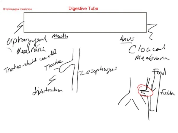
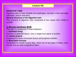
Lecture Six DIGESTIVE TUBE Digestive tube or tract includes the esophagus, stomach, small and large intestines, rectum and anus. General structure of the digestive tract The structure of digestive tube composed of four layers from inside to outside: A. Mucosa membrane (M.M) This layer is composed of three sublayers: 1. Epithelial lining Consists of epithelial tissue, vary in shape from place to another. 2. Lamina propria Composed of loose connective tissue and lymphoid nodules. 3. Muscularis mucosa Composed of smooth muscle fiber, which are of two types of fibers, inner circular and an outer longitudinal fibers.
Lecture Six B. Submucosa Consists of dense connective tissue containing blood vessels, elastic fiber. Beside that containing plexuses of nerve fibers (meissnar plexus) which are autonomic nerve fibers that control the activity of glands and the activity of muscularis mucosa. C. Muscularis externa It consist of two layers of smooth muscles, inner of circular a very much while the outer is of longitudinal arrangement. Between them plexus of nerve fibers of (Auerbach's plexus). D. Serosa or Adventitia The serosa is a thin layer of loose connective tissue, rich blood and lymph vessels and adipose tissue. This layer called serosa when connective tissue covered by mesotheial cells but called adventitia when connective tissue mergeing with connective tissue of neighboring organ. 1. Esophagus This part of the digestive tract is a muscular tube about (25cm) in length extend form pharynx to stomach. In general, it has the same layers as the rest of the digestive tract.
Lecture Six A. Mucosa membrane (M.M) 1. Epithelial lining Consists of nonkeratinized stratified squamous epithelium tissue. 2. Lamina propria Composed the loose connective tissue and less numbers of lymphoid nodules. In the lamina propria of the region near the stomach there are groups of gland, the esophageal cardiac glands, that also secrete mucus. 3. Muscularis mucosae Is composed of longitudinally organized smooth muscle only. B. Submucosa Is composed of connective tissue containing groups of small muscus-secreting glands, (the esophageal glands). C. Muscularis externa It consists of two layers of smooth muscles, inner is of circular a versed while outer is of longitudinal arrangement. At the distal end of the esophagus the muscular layer consists of only smooth muscle cells in the mid-portion, a mixture of striated and smooth muscle cells and at the proximal end only striated muscle cells. D. Adventitia Consists of loose connective tissue.
Lecture Six Stomach The stomach is a mixed exocrine-endocrine organ that digests food and secretes hormones. It is a dilated segment of the digestive tract whose main functions are to continue the digestion of carbohydrates initied in the mouth, add an acidic fluid to the ingested food transform it by muscular activity into a viscous mass (chime) and promote the initial digestion of proteins with the enzyme pepsin. It also produce a gastric lipase that digests triglycerides with the help of lingual lipase. The stomach is consisted of four regions: 1. Cardiac region Is a narrow circular band, (1.5-3cm) in width, at the transition between the esophagous and stomach. 2. Fundus Is a dom shaped evagination or projection upwards usually filled with gas. 3. Body Is the part extend form cardiac region to pyloric region and consist 2/3 of total stomach. 4. Pyloric region The part at the opining of stomach to the duodenum.
Lecture Six General structure of stomach The wall of stomach is composed 1. Mucosa membrane (M.M) When the stomach is empty the mucosa is thrown up into longitudinal folds called (Rugae) when full the rugae stretch flat and disappear. The M.M. consists of the: a. Epithelial lining Is composed simple columner epithelial tissue there are invaginations in the surface of epithelial which are called (gastric pits) in the bottom of them open brached, tubular glands called (gastric glands), their lining is simple columnar. The cells on the surface of epithelial secrete mucin called (surface mucous cells). b. Lamina propria Is composed of loose connective tissue c. Muscularis mucosa It is thinner layer composed smooth muscle fibers, inner circular fibers and outer longitudinal fibers.
Lecture Six 2. Submucosae Consist of loose connective tissue contain blood vessels and lymphatic vessels. 3. Muscularis externa Consist three layers of smooth muscle fibers, inner oblique, middle circular and outer longitudinal arrangement. 4. Serosa Consists of loose connective tissue covered the surface by mesothelium cells.
Lecture Six Types of gastric glands Gastric glands are situated in the mucosa of the membrane and are divided to the three types: 1. Cardiac glands They are found around the opening of esophagus into stomach. These are compound branched tubular gland lined by simple epithelial cells, secret mucous, the relative lengths of these glands are about 1:1 to the gastric pit. 2. Glands of fundus and the body These glands are found in the body and fundus into stomach. They are longe simple branched tubular glands. That posses four types of secretory cells: a. Mucous neck cells Simple columnar or cubodial epithelial cells secrete mucous, they are found in the neck of gland. b. Chief or zymogenic or peptic cells These simple cubodial or low columnar epi-cells, line the lower half or third of the glands. Contain microvilli and zymogen granules, this granule contain pepsinogen (inactive enzyme) pepsinogen give pepsin which is important in protein digestion. They cytoplasm appear basophilic, these cells produce lipase and amylase and carbohdryatase.
Lecture Six c. Parietal or oxyntic cells These cells are large oval or pyramidal shaped. Situated between f the chief cells the may protruded form the lateral surface of the gland. They occur singly or grouped and cytoplasma appear acidophilic. These cells contain large microvilli and produce (HCL) by secret H+ and Cl- ions and from HCl, they are also produce intrinsic factor which is necessary for absorption of B12 which is necessary for maturation of RBC. d. Argentaffin cells Present mostly at the bases of main glands between the basement membrane and zymogenic cells. They take silver stain and secrete entroglucogen which increase blood sugar level and serotonin which causes smooth muscles contraction. 3. Pyloric glands Is simple branched tubular gland that is highly coiled. They are found in the pyloric area of the stomach. Gastric pits in which these glands open, are deep funnel shaped. The length of pit is twice that of gland. These glands contain two types of cells: a. Coulmnar cells or mucosa That secret mucus it is useful to prevent the ingestion of stomach. b. Argentaffin cells Function of these cells secrete gastrin which stimulate the stomach to secret HCl and pepsine.