Download
1 / 15
150 likes | 166 Vues
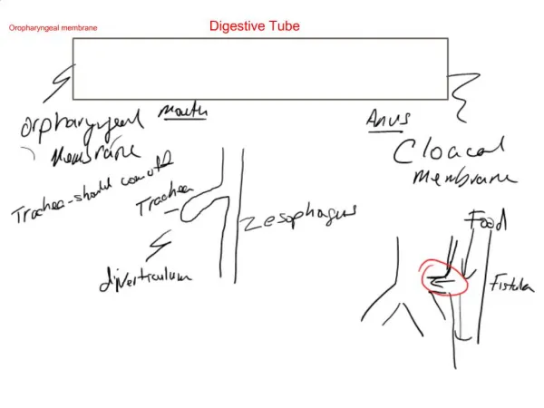
The digestive system. The digestive system consists of :- A- Oral cavity B- Digestive tube C- Digestive glands. A-oral cavity Includes the lips , cheeks , tongue , teeth , pharynx , palate , and salivary glands . lips

E N D
The digestive system The digestive system consists of :- A- Oral cavity B- Digestive tube C- Digestive glands
A-oral cavity Includes the lips , cheeks , tongue , teeth , pharynx , palate , and salivary glands . lips are consists of strained muscles fibers called ( orbicularis oris ) embedded in connective tissue. The lips covered from outside by skin contain hair follicle, sebaceous and sweat glands. While internal side covered by mucous membrane (m.m) this membrane consist of non keratinized squamous epithelial tissue resting on lamina propria which consists of loose connective tissue containing some mucous glands called ( labial glands). The margin of the lip is transitional from between the skin and the mucous membrane have large numbers of capillaries therefore it have a red color . The epidermis is not heavily keratininzed it contain large amounts of Elidin which is caused it translucent . • the function of this system are digestion and absorption of food molecules into blood and lymphatic . the molecules necessary for the maintenance , growth and energy needs of the body .
2-Cheeks • They are like lips , consist of skeletal muscles called (Buccinator) • that is covered by skin from outside and from inside by mucous • membrane . • The (m.m) is connected to the muscle by elastic fibers called • ( mucosal fold ) epithelial tissue rich supplied with capillaries. • 3-Palate • can be divided in two types :- • A-Hared palate. • Composed of keratinized stratified squamous epithelial tissue , there is • no submucosa and the epithelial tissue directly connected firmaly to • the periosteum of the bone . • B-Soft palate. • Is lined by nonkeratinized stratified squamous epithelial tissue , there is wide submucous between epithelial tissue and the periosteum. • 4-Tongue • is a muscular organ on the floor of the oral cavity .its functions:- • 1-play important role in the formation of speech sounds. • 2-In processing food material between teeth during mastigation. • 3-The sense of the taste is also located mainly in the tongue . • The tongue is consist of a mass of skeletal muscles which covered by mucosa membrane . the muscles fibers cross on another orientation in all direction that we can move tongue in all direction.
The tongue muscles are covered by mucous membrane this membrane of the upper surface is divided in anterior part and posterior part by (V- shaped) sulcus called terminal sulcus . the anterior part of the tongue include 2/3 it called oral part of the body , while the pharyngeal root include 1/3 (posterior part). • Also found longitudinal sulcus divides the tongue to half parts called medium sulcus.
The mucosa membrane of upper surface of the oral part of the body contain numerous projections called ( papillae). • The papillae are divided into four types :- • 1-Filiform papillae • These are the most numerous , and are distributed over the whole surface • of the tongue . each papilla has a thin core of connective tissue and is • covered by keratinized stratified epithelial tissue resting on the lamina • propria and has no taste bunds . • 2-Fugiform papillae • These are fewer and larger than the filiform papillae and are scattered irregularly among them all over the surface of the tongue . • They have mushroom shape . it is covered by semi keratinized stratified epithelial having blood vessels in the lamina propria and have • some taste bund in their epithelium. • 3-Circumvallate papillae • Their number from (nine fifteen ), are much larger than the fungiform papillae , and are arranged along the terminal sulcus . it is covered • by non-keratinized stratified epithelial tissue , lamina propria containing blood vessels . A deep furrow surrounds the base of each papilla. • Open at the base of the circular furrows serous (Von Ebner,s) glands. Their duct opens mainly into the circular sulcus surrounding the • circumvallate papillae. Taste buds are present on the side walls of there papillae .
4-Foliate papillae • These are poorly developed in humans . consist of parallel low ridges separated by deep mucosal clefts . they occur on the lateral edge of the tongue in aged humans the foliate papillae may not be recognized . in younger individuals they are easily found on the posterior lateral surface of the tongue and contain many taste buds in the epithelium of the walls of neighboring papillae small serious glands empty into the clefts. In some animals such as the rabbit , foliate papillae constitute the principal site of aggregation of taste buds. • The root of the tongue appears irregular in shaped of the presence of lingual tonsils . the mucosa membrane covering the root is smooth because it has no papillae . • Test buds • Taste buds present in fungiform , foliate and circumvallate papillae . they are barrel shaped . these buds containing two pores (gustatory and nerve pore ) , and two types of cells are present :-
1-Gustatory cells or neuro epithelial taste cells . • These are slender bipolar cells with a central enlargement for the nucleus they have synaptic vesicles which have sensory nerveendings. • 2-Sustentacular or supporting cells • These are long and with broad oval nuclei supporting the gustatory and forming the walls of the taste bud .
5-Teeth • Teeth are major component of the oral cavity and are essential for the digestive process. Teeth are embedded in and attached to the maxilla and mandible. In adult humansthere are normally 32 permanent teeth . each tooth is composed of a portion that projects above the gingival the ( called crown) and one or more roots below the gingiva. These two parts meet at the neck each tooth contain the pulp cavity . the pulp cavity. the pulp cavity extends to the apex of the root . • Tooth consists of two parts of tissues :- • Hard tissues in the teeth . • This is divided into :- • 1-Dentin • Dentin is a calcified tissue like borne but harder , because of its higher content of calcium salts (70 % of dry weight ). • It is composed mainly of type 1 collagen fibrils,glycosminglycans and calcium salts in the for of hydroxyapatite crystals. The organic matrix of dentin is secreted by odontoblasts (cells that line the internal surface of the tooth ) separating it from the pulp cavity. The odontoblast is a slender polarized cell , that produces organic matrix only at the dentinal surface . • 2-Enamel • Is the hardest component of the human body and it is the richest in calcium it consist of about ( 95% calcium salts) mainly hydroxyapatite , (5% organic material ) and water . Enamel is produced by cells of ectodermal origin , whereas most of the other structures by cells of ectodermal origin , whereas most of the other structures of teeth derive from mesodermal or neural crest cells. • The organic enamel matrix is not composed of collagen fibrils but of at least two heterogenous classes of proteins called amelogenins and enamelins .
Enamel consists of elongated rods or columns of hydroxyyapatie crystals called ( enamel rods or prisms). • Enamel matrix is secreted by cells called ameloblasts. These are tall columnar cells posses numerous mitochondria in the region below the nucleus . • The dentin of the crown of teeth covered by the extremely hard enamel. • 3-Cementum • This tissue covers the dentin of the root and is similar in composition to bone although haversion and blood vessels are absent. It is thicker in the apical region of the root , where there are cementocytes like osteocytes , they are encased in lacunae that communicate through canaliculi like bone tissue . • B-Soft tissues in the teeth divided into :- • 1-Pulp • Tooth pulp consists of loose connective tissue . its main components are odontoblasts fibroblasts , thin collagen fibrils and a ground substance that contains glycosaminoglycans . • Pulp is a highly innervated and vascularized tissue blood vessels and myelinated nerve fibers enter the apical foramen and divide into numerous branches . some nerve fibers lose their myelin sheaths and extend for a short distance into the dentinal tubeless . these fibers are sensitive to pain . • 2-Periodontal ligament • The periodontal ligament is composed of a special type of dense connective tissue and loose . the dense connective tissue contains collagen fibers and fibroblasts are arranged parallel to the long axis of the collagen fibers.
The loose connective tissue in the periodontal ligament contains B.V. and nerve endings in addition to the cells and thin collagenous fibers . • The periodontal ligament provides for :- • Attachment • Support • Nutrition of adjacent structures • 2-Gingiva • The gingiva is a mucous membrane firmly to the periosteum of the maxillary and mandibular bones it is composed of stratified sguamous epithelial tissue and numerous connective tissue papillae.