Download
1 / 50
530 likes | 847 Vues
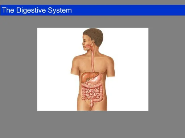
LECTURE. DIGESTIVE SYSTEM part 1 Oral cavity, esophagus & stomach. Department of histology, cytology and embryology KhNMU. General description and significance. The digestive tract is: a long tube extending from oral cavity to the anus and associated glands .

E N D
LECTURE DIGESTIVE SYSTEMpart 1Oral cavity, esophagus & stomach Department of histology, cytology and embryology KhNMU
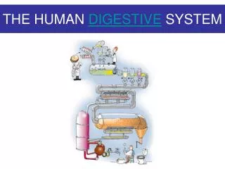
General description and significance • The digestive tract is: • a long tube extending from oral cavity to the anus and • associated glands. • Main functions are: ingestion, fragmentation, digestion, absorption of nutrients and elimination of waste products.
Classification and embryogenesis 3 compartments: • Anterior Oral cavity, pharynx, esophagus • Middle Gastrointestinal tract • Posterior Last 1/3 rectum, anus
Embryogenesis Gut – from endoderm, stomatodaeum, proctodaeum – from ectoderm Connective tissue, muscles are from mesoderm amnion yolk sac
Embryogenesis foregut hindgut midgut Cloacal Plate Oral Plate Allantois Heart • Yolk Sac
General plan of structure • FOUR principal membranes: • Mucosa: Epithelium Lamina propria Muscularismucosae • Submucosa • Muscularisexterna • Serosa or adventitia
3a 1a 2a 1b 1c 2 2b 4 3b
Mucosa:2. lamina propria – con.t. – nutrition, support, protection (GALT), absorption, secrition (glands), villi 3. muscularis mucosae • Submucosa - con.t.- big bl.vessels, glands, nerve plexuses, lymphatic nodules, • provides motility of mucosa • Muscularisexterna – circular, longitudinal layers • Serosa or adventitia
Oral Cavity consistsofanumberofsuborgans Functions: Ingestion, STRUCTURE: Fragmentation 1. Mucosa Moistening 2. Submucosa Speech (not always present ! ) Facial expression 3. Muscularis ext. Sensory reception (facial muscles) Breathing 4. Adventitia (absent)
skin side skin oralside oral ss ts • Lip ts lp bv sk The outer surface (skin) of the lip is covered by thin skin (ts). The inner surface (oral) is lined by a mucosa The transitional zone sg ss sk sk low mag.
ss Lip red area Vermilion - “red” transitionalzone In newborn there are small villi for sucking (Krok). Thecheekissimilartothelip. p bv
f Tongue C E Epithelium (E) & underlying ct. Skeletal muscle (SM) runs in 3 planes. Embedded in the skeletal muscle lie glands (G). Papillae. Most numerous are conical, keratinizedfiliform (f) papillae; function – general sensation fungiformand foliatepapillae are scattered. At the sulcus terminalis, lie 8 -12 large circumvallate papillae (C). - Taste sensation SM G
Tongue, filiform & fungiform papillae ss ss fungi fili nss fili ct ct Circumvallate papilla
Tongue • papillawithtastebuds fp high tb med
Maxilla unerupted tooth Hard Palate b hard palate epithelium • The mucosa (m) is tightly bound to the bone (b). The epithelium (ss) is disturbed in chewing and swallowing and so tends to be keratinized, and lamina propria forms deep papillae, protruding in the epithelium. ss m low med
Soft palate Epithelium sm sg Itsuppersurfacefacestherespiratorypassages- pseudostratifiedepithelium. Facing oral cavity epithelium isstratifiedsquamous. Krok ! oral cavity
c ss Lingual Tonsil T mg Lingual tonsil (T) locates on the dorsum of the tongue. Together with palatine, pharyngeal tonsils form "ring". At time of chronic inflammation may undergo tonsilectomy. sk
Toothdevelopment • Enamel organ (epith) • Ameloblasts • Enamel • Dental papilla • Odontoblasts • Dentine • Dental sac –cementum, pulp
SALIVARY GLANDS • Saliva: water, mucus, amylase, lysozyme, a/b, ions • I. Large salivary glands 3 pairs: parotid, submandibular, sublingual function - IN RESPONce TOPARASYMPATHETIC ACTIVITY • II. Minor salivary glandsfunction - CONTINUOUSLY
General structure • Compound branched acinar or acino-tubular glands • Connective tissue capsule • Lobulated structure • Lobules contain secretory units and small ducts • Interlobular connective tissue with ducts and vessels
5 Intercalated duct Serous acinus Striated duct, Next are interlobular and general Mucousacinus
Secretory unit Myoepithelial cells (contractile) surround secretory portions and small ducts – intercalated and striated Nucleus Myoepithelial cells
su sd Parotid The parotid is a serous gland secreting amylase. Secretory unit (su) is acinar. Pyramidal acinar cells have round basally-placed nuclei (n) with abundant basal RER RER n high low low
itd Sublingual gland id This gland has mucous & mixedsecretory units but mostly mucous. med me sd d msu low id me n high • Mixedacinuswitha • Serousdemilune msu
lobule Submandibular gland m/s It is predominantlyserousglandshowsblue, pureserousacini (sa) & pale, mucousand mixed with serousdemilunes (m/s). low id ed sa high med
Intercalated duct Demilune demilune Parotid gland sublingual gland Sublingual gland Compare! Striated duct Demilune Striated duct Submandibular gland
Esophagus (=Pharynx) 6-7 longitudinal folds. The muscularisexterna. The upper 1/3rd is composed of skeletal muscle; the lower 1/3rd - smooth muscle; the middle 1/3rd shows a blending of the two varieties of muscle. Epithelium is stratified squamous nonkeratinized
Esophagus • 1) Mucosa-epithelium, lamina • propria & muscularis mucosae, • 2) Submucosa, • 3) Muscularis • externa • 4) Adventitia
Esophagus ss mg • Mucosal cardiac glands (mg) are in the lamina propria (lp) (mm) in the upper and lower thirds of the esophagus. • If they are not fully effective, the excessive reflux results in pyrosis (heartburn) – the rise of the gastric contents upward toward the neck. lp sm mm me
Esophagus mm mg A mucousesophageal glands proper (mg) lieinthesubmucosa (s). s me
Esophagus, • middle 1/3rd - med. mag. • . m L sub muscularisexterna (me). = smooth(sm) & skeletalmuscle (sk). lymphatic nodules (L)areinthemucosa (m). sm me sk
submucosal gl. Esophagus med lymphatic nodule M' high The enteric nervous system is extensive and its neurons and fibersform in thesubmucosa (Meissner’s plexus) (M') and in the muscularis externa (Auerbach’s plexus) (A'). lumen str.sq.epi musc. muc. A' high muscul. externa. submuc.
E 1 S gp sc 2 low sc 3 E S gp med high • Esophagus/Stomach junction. - • Epithelium is changed from stratified squamous to • simple columnar (sc).
Stomach - general • Theprocessofdigestionessentiallybeginsinthestomach; littleabsorptionand excretion also occurhere. • Thestomachiscomposedofamucosa, submucosa, muscularisexterna & serosa. • Themucosalliningisasimplecolumnarsecretoryepithelium (mucous). • Folds (rugae),gastricpits, mammilated areas. • Glandsvaryindifferentregionsofthestomach.
Stomach mucosa: ```epithelium lamina propria:gastric glands; muscularismucosae: both circular and longitudinal layersof SM. submucosa: muscularis: the inner oblique layer, circular layer and longitudinal layer of SM. serosa
sc gp • Stomach, mucosal lining & gastric pits Gastric pits (gp) are invaginations of a simple columnar epithelium The mucus blanket which protects the lining from stomach acids is present here.
Lamina propria contains glands • fundic glands (in the body, fundus) secrete the enzymes and acid of the stomach. While cardiacand pyloricglands predominantly secrete mucus. • The fundic glands are simple tubular. • They contain 4 cell types:
1. Mucous neck cells2. Chief cells , or zymogenic cells • columnar and basophilic cells - in the body of the glands. • produce pepsinogen and lipase. 3. Parietal cells secrete hydrochloric acid ( HCL ) and intrinsic factor. The latter is necessary for absorption of vitamin B12 in the ileum
4. Enteroendocrine cells (APUD) • 20 different types • 4 principal hormones: • GastrinHcl secretion • Secretin, cholecystokinin (CCK), gastric inhibitory peptide (GIP) pancreatic and gallblader activity and gastric secretion
Pernicious anemiafollows the absence of parietal cells (i.e. loss of epithelium due to gastric ulcer) • Disturbance of different cells may call the different pathology. • Digestion of different substances begins in different regions.
r gp Fundic stomach m sm me fg low sm 2 mm Thelowestmag. showsthemucosa(m), submucosa(sm) withfoldscalledrugae(r) & themuscularisexterna (me). Athighermag. gastricpits (gp), fundicglands (fg), &muscularismucosa (mm) comprisesthemucosa. Outsideisthesubmucosa (sm) andmuscularisexterna (me). me med
Fundic stomachmucosa gastric pit Isthmus Neck Base Infundicstomach,gastricpitsoccupyabout 1/5 themucosa; fundicglandscomprisetheremaining 3/4ths. fundic gland
sc gastric pit • CardiacStomach, Gastricpits occupy ½ of mucosa. Gastricglandsare simple tubular and slightly branched Cells are mucus-secreting and occational endocrine cardiac glands mm
Stomach, pyloric gastric pit gastric gland Gastric pits occupy 3/4ths the depth of the mucosa; the remaining 1/4th are pyloric glands – short and branched. The major cell of the gland is the pale mucous (mn); parietal cells are absent as the pyloric stomach nears the intestine. mn low
smooth muscle outer inner m • FundicStomach, nervesupply, - med & highmag Elementsofthesubmucosal (s) andmyenteric (m) nerveplexusesarepresent. middle fundic glands submucosa muscularis externa artery musc. mucosa s vein