Download
1 / 25
250 likes | 391 Vues
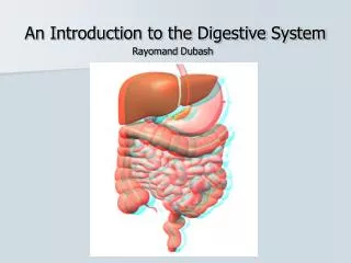
INTRODUCTION TO DIGESTIVE SYSTEM ANATOMY. 12. November . 2013 Tuesday. Kaan Yücel M.D., Ph.D . .INTRODUCTION. Food passes from the mouth and pharynx esophagus stomach mixes with gastric secretions. Digestion mostly occurs in the stomach and duodenum . Peristalsis.

E N D
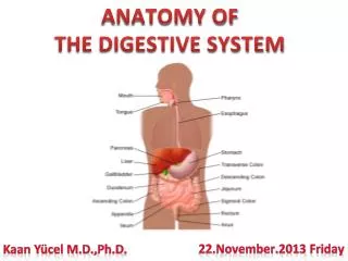
INTRODUCTION TO DIGESTIVE SYSTEM ANATOMY 12.November.2013Tuesday • Kaan Yücel M.D., Ph.D.
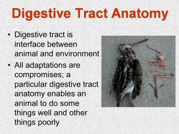
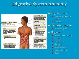
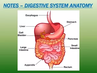
.INTRODUCTION Food passes from the mouth and pharynx esophagus stomach mixes with gastric secretions. Digestion mostly occurs in the stomach and duodenum.
Peristalsis a series of ring-like contraction waves begins around the middle of the stomach moves slowly toward the pylorus • responsible for • mixing the masticated (chewed) food mass with gastric juices • emptying the contents of the stomach into the duodenum.
Absorption Absorption of chemical compounds occurs principally in the small intestine a coiled 5- to 6-m-long tube duodenum, jejunum, and ileum.
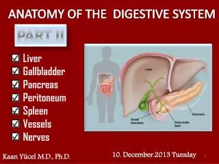
Peristalsis also occurs in the jejunum and ileum. is not forceful unless an obstruction is present. Stomach continuous with duodenum receives the openings of the ducts from pancreas & livermajor glands of the digestive tract.
Largeintestine Cecumreceives the terminal part of the ileum Appendix Colon(ascending, transverse, descending, and sigmoid) Rectum Anal canal Most reabsorption of water occurs in the ascending colon. Feces form in the descending and sigmoid colon accumulate in therectumbefore defecation.
2. SKELATAL ANATOMY OF THE ORAL & NECK REGIONS Oralcavity inferior to the nasal cavities. has a roof and floor, and lateral walls. opens onto the face through the oral fissure. continuous with the cavity of the pharynx @ oropharyngealisthmus.
2. SKELATAL ANATOMY OF THE ORAL REGION Bones contribute to the skeletal framework of the oral cavity paired Maxillae Palatine bone Temporal bones unpaired Mandible Sphenoid Hyoid bone
2. SKELATAL ANATOMY OF THE ORAL REGION cartilaginous parts of the pharyngotympanic tubes on the inferior aspect of the base of the skull related to the attachment of muscles of the soft palate.
2. SKELATAL ANATOMY OF THE ORAL REGION The styloid process and inferior aspect of the petrous part of the temporal bone provide attachment for muscles associated with the tongue and soft palate, respectively.
2. SKELATAL ANATOMY OF THE ORAL REGION Mandible bone of the lower jaw. a body of right and left parts, fused anteriorly in the midline and two rami. The hyoid bone is a small U-shaped bone in the neck between the larynx and the mandible.
2. NECK a tube providing continuity from the head to the trunk extends anteriorly lower border of the mandible upper surface of the manubrium of sternum posteriorly superior nuchal line on occipital boneintervertebral disc between the CVII & TI.
3. SKELATAL ANATOMY OF ABDOMINAL REGION Abdomen a roughly cylindrical chamber inferior margin of the thorax superior margin of the pelvis and the lower limb.
3. SKELATAL ANATOMY OF ABDOMINAL REGION Sup. opening of the abdomen: inferior thoracic aperture closed by the diaphragm. Inferiorly, the deep abdominal wall is continuous with the pelvic wall at the pelvic inlet. Superficially, the inferior limit of the abdominal wall is the superior margin of the lower limb.
4. SKELATAL ANATOMY OF THE PELVIC REGION • Pelvisis divided into two regions: • Falsepelvis (Greaterpelvis) • superior region related to upper parts of the pelvic bones& lower lumbar vertebrae • generally considered part of the abdomen • True pelvis (Lesserpelvis) • related to the inferior parts of the pelvic bones, sacrum, and coccyx • has an inlet and an outlet.
4. SKELATAL ANATOMY OF THE PELVIC REGION The bowl-shaped pelvic cavity enclosed by the true pelvis consists of the pelvic inlet, walls, and floor. continuous superiorly with abdominal cavity contains elements of the urinary, gastrointestinal, and reproductive systems.
4. SKELATAL ANATOMY OF THE PELVIC REGION Pelvicinlet somewhat heart shaped completely ringed by bone. Posteriorly, the inlet is bordered by the body of vertebra SI. pelvic outlet diamond-shaped formed by both bone and ligaments. limited anteriorly in the midline by pubic symphysis.
4. SKELATAL ANATOMY OF THE PELVIC REGION The bones of the pelvis right and left pelvic (hip) bones Sacrum Coccyx
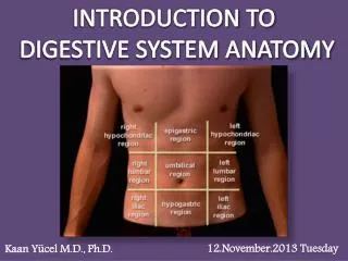
5. ABDOMINAL REGIONS Visualization of the position of abdominal viscera is fundamental to a physical examination. Some of these viscera or their parts can be felt by palpating through the abdominal wall. Topographical divisions of the abdomen are used to describe the location of abdominal organs and the pain associated with abdominal problems.
FOUR-QUADRANT PATTERN A horizontal transumbilical plane passing through the umbilicus & intervertebral disc between vertebrae LIII and LIV intersecting with the vertical median plane • right upper • left upper • right lower • left lower quadrants
NINE-REGION PATTERN • 2 horizontal PLANES • Superiorhorizonalplane (Subcostalplane) • immediately inferior to the costal margins, • at the lower border of the costal cartilage of rib X • passes posteriorly through the body of vertebra LIII.
NINE-REGION PATTERN • 2 horizontal PLANES • Inferiorhorizonalplane(Intertubercularplane) • connects the tubercles of the iliac crests • palpable structures 5 cm posterior to the anterior superior iliac spines • passes through the upper part of the body of vertebra LV.
NINE-REGION PATTERN • 2 horizontal PLANES • Inferiorhorizonalplane(Intertubercularplane) • connects the tubercles of the iliac crests • palpable structures 5 cm posterior to the anterior superior iliac spines • passes through the upper part of the body of vertebra LV.
NINE-REGION PATTERN 2 VERTICAL PLANES • pass from the midpoint of the clavicles inferiorly to a point • midway between • anterior superior iliac spine • & • pubic symphysis
NINE-REGION PATTERN • Superiorly • right hypochondrium • epigastric region • left hypochondrium • Inferiorly • right groin (inguinal region) • pubic region • left groin (inguinal region) • In the middle • right flank (lateral region) • umbilical region • left flank (lateral region)