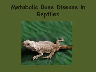
Metabolic Bone Disorders
730 likes | 1.13k Vues
Metabolic Bone Disorders. Objectives. Differentiate metabolic bone disorders by etiology, treatment and outcome. Outline common nursing diagnoses, outcome criteria and interventions for common metabolic bone disorders. Bone Cell Types. Osteoblast Forms bone & mineralization of matrix

Metabolic Bone Disorders
E N D
Presentation Transcript
Objectives • Differentiate metabolic bone disorders by etiology, treatment and outcome. • Outline common nursing diagnoses, outcome criteria and interventions for common metabolic bone disorders.
Bone Cell Types • Osteoblast • Forms bone & mineralization of matrix • Osteocyte • Transformed osteoblast • Maintains bone found in matrix • Osteoclast • Breaks down bone salts • Responsible for bone reabsorption
Osteoblasts “Baby bone cells” “Building Blocks” Osteoclasts “Clean up” cells Osteocytes “Cycle” of bone Bone Cell Mnemonics
Question #1: Which statement is true of osteoblasts? a. They transform osteocytes into osteoblasts. b. They maintain cells within the bone matrix. c. Osteoblasts form bone cells within matrix. d. Osteoblasts break down bone salts.
Answer #1. Which statement is true of Osteoblasts? c. Osteoblasts form bone cells within matrix. Rationale: Osteoblasts are “bone builders”; the other responses are related to functions of other bone cell types.
Hormonal Regulationof Bone Metabolism • Thyroid gland • Thyroxine, triodothronine & calcitonin • Regulated by TSH / TRH & calcitonin by plasma levels of calcium • Parathyroid gland • Parathormone PTH (protein hormone) • Regulated by serum ionized calcium levels
Hormonal Regulationof Bone Metabolism • Anterior pituitary gland • ACTH / TSH / FSH / LH / Prolactin • Regulated by hypothalamus • Adrenal cortex • Glucocortcoids / mineralcorticoids & androgens • Estrogen • Increased osteoblast activity • Retention of calcium and phosphate
Question #2: Which hormone is the most important for regulating serum calcium levels because it acts directly on bone and kidneys? a. Parathyroid hormone. b. Growth hormone. c. Calcitonin. d. Adrenal corticosteroids.
Answer #2: Which hormone is the most important for regulating serum calcium levels because it acts directly on bone and kidneys? a. Parathyroid Hormone. Rationale: As noted earlier, this hormone acts directly on bone and kidneys
Hyperparathyroidism • Mainly two types • Primary- cause unknown but thought to be familial and characterized by excessive secretion of PTH • Secondary-usually due to disease state such as renal failure which causes decrease in ionized serum calcium levels • Excess Secretion of PTH • Interrupts metabolism of calcium / phosphate / Bone
Hyperparathyroidism- Pathophysiology • Although primary/secondary cause either hypo or hypercalcemia, end result remains elevated levels of PTH which causes eventual hypercalcemia and multisystem problems
Primary Results in: Hypercalcemia Causes Adenoma / Carcinoma Genetic / Multiple Endocrine Disorder Secondary Results in initial hypocalcemia followed by hypercalcemia Causes Chronic Renal Failure / Malabsorption Syndromes / Vitamin D Deficiency Hyperparathyroidism
Hyperparathyroidism • Clinical manifestations • Bones – Demineralization due to excessive osteoclast and osteocyte activity • Kidneys – renal calculi, UTI • GI– Anorexia / NV, pancreatitis, peptic ulcers, constipation, hypergastrinemia • Psychiatric issues • Muscle weakness, myalgias
Hyperparathyroidism • Diagnostics • All other causes of hypercalcemia must be eliminated first • 6 month history of symptoms of hypercalcemia • Kidney stones, hypophosphatemia, hypochloremia • Serum Calcium Levels - >10mg/dl • PTH Assay – ↑1° • Radioactive Iodine Uptake Test - ↓ • Subclinical / Post- Partum / Acute Thyroiditis • Urinary Calcium – ↑(24 Hr Specimen) • DEXA Bone Density - ↓
Hyperparathyroidism • Clinical Management • Adequate Hydration • Increase urinary excretion of Ca++ with diuretics • Drugs that decrease resorption of Ca++ by bone-biphosphates, calcitonin • Surgery • Parathyroidectomy – NOT Often Recommended • Leaves ½ of one Lobe of the Parathyroid • Remove Adenoma
Question #3: Ms. Jones is a 60-year-old female who presents in the Clinic with a 6 month history of frequent renal stones, abdominal pain, muscle aches and several fractures of her metatarsals. The nurse would suspect: a. Gout. b. Hyperparathyroidism. c. Hypoparathyroidism. d. Paget’s Disease.
Answer # 3: Ms. Jones is a 60-year-old female who presents in the Clinic with a 6 month history of frequent renal stones, abdominal pain, muscle aches and several fractures of her metatarsals. The nurse would suspect: b. Hyperparathyroidism. Rationale: As defined earlier, these are common s/s of hyperparathyroidism
Question #4: In order to confirm this diagnosis, diagnostic testing needs to be performed. As the Nurse you know: a. That you can rely on one blood sample to give complete results. b. The patient will need blood work, DEXA scans, and 24 hour urine samples c. That you can rely on urine testing alone. d. The tests will most likely be inconclusive.
Answer #4: In order to confirm this diagnosis, diagnostic testing needs to be performed. As the Nurse you know: b. You will need to have results of serum Ca++, phosphate, magnesium, bicarbonate levels as well as a DEXA scan and a 24 hour urine for excreted Ca++ Rationale: DEXA scan shows demineralization of bone, 24 hour urine shows excess Ca++, and abnormal serum levels of trace elements
Question #5: Mrs. Jones is diagnosed with hyperparathyroidism. As the nurse doing the patient teaching, you are aware that adequate hydration is essential in preventing: a. Constipation. b. Hypercalcemia. c. Alteration in fluid balance. d. All of the above.
Answer #5: Mrs. Jones is diagnosed with Hyperparathyroidism. As the nurse doing the patient teaching, You are aware that adequate hydration is essential in preventing: d. All of the above. Rationale: Adequate hydrationhelps to prevent constipation, hypercalcemia and fluid balance alterations
Hypoparathyroidism • Decreased Secretion of PTH • Most commonly caused by injury to parathyroid gland during surgery • Can also be caused by hypomagnesemia • Pathophysiology • Bones – Mineralization Bone Resorption • Hypocalcemia / Intestinal Ca+ Absorption • Metabolic Alkalosis (Mild) • Parkinson-like Symptoms
Hypoparathyroidism • Clinical Presentation • Mental Fatigue • Abdominal Pain • Patient History of Alcoholism • Physical Examination • Muscle Spasm / Tetany / Excitability • Deep Tendon Reflexes • Dry Skin / Hair Loss / • Weakened Tooth Enamel
Hypoparathyroidism • Diagnostics • Serum Calcium Levels – DECREASED • Serum Phosphorus – INCREASED • Low Vitamin D Levels • Urinary Calcium –DECREASED • X-Rays • Increased Bone Density
Hypoparathyroidism • Clinical Management • Acute condition • MEDICAL EMERGENCY • Prevent larygneal spasms- administer IV Ca++ gluconate/carbonate STAT! • Chronic condition • Lifetime Vitamin D therapy • Calcium supplementation- 1 to 3 gm/day • Muscle relaxants to control muscular spasms • Drugs to reduce GI absorption of phosphorous
Osteomalacia (Adult Rickets) • Inadequate and delayed mineralization of osteoid in mature compact and spongy bone • Major deficit is in Vitamin D , which is required for Ca++ uptake in intestines • Decreased Ca++ stimulates PTH, which does increase Ca++, but also increases phosphate excretion by kidney • When phosphate levels too low, mineralization cannot occur
Osteomalacia (Adult Rickets) con’t • Etiology • More prevalent in extreme preemies, elderly, those following strict macrobiotic vegetarian diets and persons on anticonvulsant Rx • Pancreatic insufficiency • Hepatobiliary diseases • Lack of bile salts decreases absorption of Vit D • Malabsorption syndromes • Hyperthyroidism • Rare in US due to fortification of foods • Common in GB and Middle Eastern Countries
Osteomalacia • Clinical Presentation • Generalized body aches /LBP as well as hip pain • Lower extremity pain & deformity • Physical examination • Scoliosis / kyphosis of spine • Deformities of weight bearing bones • Muscle weakness leading to classic waddling gait • Generalized Malaise
Osteomalacia • Diagnostics • Serum Ca++ –↓ or Normal • Serum inorganic Phosphate ↑> 5.5 • Vitamin D ↓ • BUN & creatinine ↑ • Alkaline Phosphatase & PTH ↑ • Bone bx to determine aluminum levels • X-Rays • Demineralization • Pseudofractures • Bowing of long bones
Osteomalacia • Clinical Management • Correcting serum Ca++ & phosphorous • Chelating bone aluminum if needed • Suppressing hyperthyroidism • Supplement with Vitamin D • Administer Ca++ carbonate to ↓ hyperphosphatemia • Renal dialysis/transplant for renal osteodystrophy • Correction of associated intestinal disorders
Question #6: X-rays of a patient with Osteomalacia would reveal: a. Increased bone density. b. Stress fractures. c. Normal joint alignment. d. Demineralization.
Answer #6: X-rays of a patient with Osteomalacia would reveal: d. Demineralization. Rationale: As calcium and phosphorus levels are decreased, demineralization can be noted on x-ray
Osteoporosis • Most common metabolic bone disease • Reduction of bone mass density (BMD) fractures • Estrogen deficiency leads to a rapid in BMD • Rapid bone loss may occur • Up to 20% during the first 5-7 years post-menopause • Surgically induced menopause • Results in severe decrease in BMD regardless of age
Inherited Gender / Ethnicity Body composition Gyn considerations Family History Hx. Of osteoporosis Medical Conditions Rheumatoid arthritis Thyroid / Liver Dz Spinal cord injury Behavioral Physical activity level Nutritional status Lifestyle habits Medications Thyroid replacement Corticosteroid use Antacids Long term anti-convulsant use Osteoporosis – Risk Factors
Osteoporosis • Clinical Presentation • Attire Ill fitting clothes • Height Recent loss of height • Spine (Posture) Kyphosis • Chest/ Abdomen Chest resting on protruding abdomen • Gait Slow reciprocal – Wide base stance
Osteoporosis • Differential Diagnosis • Urinary calcium - ↑ in secondary osteoporosis • Biochemical markers of bone resorption • Urinary pyridinoline- ↑for a variety of metabolic bone diseases • X-Rays • ↑ density often not seen until 50% loss • DEXA • Hip / Lumbosacral spine -↑
Osteoporosis – Fracture Risk • Essential to ALL groups • Post-menopausal & elderly MOST at risk for fracture • Bone strength depends on • Mass • Architecture • Bone Quality • BMD Testing • Bone Mass Measurement Act
Osteoporosis • Nutritional support • Calcium intake levels • RDA based on age • Co-Factors • Vitamin D • Serum 1,25-dihydroxyvitamin D3 • Exercise • Weight bearing exercise 2-3 x week
Anti-Resorptive Medication • Estrogen • Prevents bone resorption • Most commonly used • Start within 3 Yrs of menopause • Positive effect of calcium absorption & calcitonin • risk of endometrial cancer – progesterone MUST be added if no hysterectomy • Oral / Transdermal • New data shows no change in CV risk
Anti-Resorptive Medication • Calcitonin • Inhibits osteoclasts – prevents bone resorption • Tx. postmenopausal osteoporosis • Males & females • In conjunction with calcium & Vitamin D • Analgesic properties • Intranasal administration
Anti-Resorptive Medication • Bisphosphonates Non-Hormonal agent • Highly selective osteoclast inhibitor • Indicated for treatment & prevention & osteoporosis in men • BMD 2 standard dev. below norm for young adults • SE – GI disorders / Esophageal & gastric ulcers
Anti-Resorptive Medication • SERM - Selective Estrogen Receptor Modulator • Indicated for prevention • Enhances beneficial effects of estrogen without increasing risks to breast / uterus • Caution use in patients at risk for DVT
Bone Forming Agents • Slow-Release calcium fluoride • Stimulate osteoblast activity • New bone matrix remains brittle • Not effective with severe demineralization • Must have adequate calcium intake See Handout for medications
Osteoporosis • Surgical intervention for vertebral fractures • Vertebroplasty • High pressure injection of bone cement through pedicles to vertebral body • Contraindicated in severe vertebral body collapse
Osteoporosis • Surgical intervention for vertebral fractures • Kyphoplasty • Bone tamp through cortical window • Inflation of bladder in vertebral body • Injection of bone cement under LOW PRESSURE
Osteoporosis • Physiological • Decreased respiratory function • Kyphotic deformity • GI/Bowel alteration • Protrusion of abdomen • Medications • Self-care deficits