Download
1 / 27
400 likes | 1.2k Vues
Tracheoesophageal Fistula with Esophageal Atresia Clinical Case Presentation. Presented by Magidah Kobty RNC, SNNP GNRS 5632 May 27, 2014. Objectives . Maternal History Maternal and Fetal Risks and Complications Delivery and Stabilization Admission exam and diagnostics

E N D
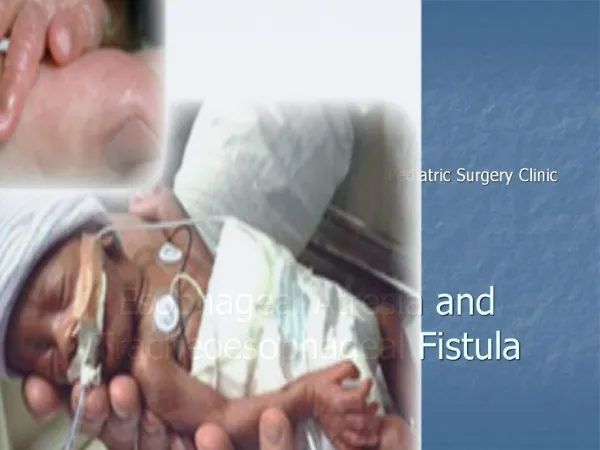
Tracheoesophageal Fistula with Esophageal Atresia Clinical Case Presentation Presented by Magidah Kobty RNC, SNNP GNRS 5632 May 27, 2014
Objectives • Maternal History • Maternal and Fetal Risks and Complications • Delivery and Stabilization • Admission exam and diagnostics • Primary admission diagnoses • Etiology and Pathophysiology of Primary Admission Diagnoses • Initial Plan of Care • Hospital Course by Symptoms • Medications • Pertinent Theories and Evidence Based Practice • Family Interactions • Discharge Plan and Follow Up Magidah Kobty RNC SNNP
Maternal History • 37 year old White G 3, P 2, Pr 0, LC 2 • Good prenatal care • Estimated date of delivery: 05/17/2014 • Maternal Blood Type: O Positive • Gestational Diabetes • Maternal Labs • Hepatitis B Negative • HIV Negative • Rubella Immune • RPR non reactive • GBS negative Magidah Kobty RNC SNNP
Maternal & Fetal Risks and Complications • Advanced Maternal Age • >35 yrs old • Increased risk of gestational diabetes • Increased risk of miscarriage or chromosomal abnormality • Gestational Diabetes • Occurs in 7% of pregnancies • Maternal Risks • High blood pressure and preeclampsia • Urinary tract infection • Future risk of developing Type 2 diabetes risk – non gestational • Risks to infant • Preterm delivery and RDS • Hypoglycemia • LGA infants, shoulder dystocia, birth injury • Increased risk of Type 2 Diabetes later in life • No fetal risks identified prenatally (Mills, 2011) Magidah Kobty RNC SNNP (Ryan, 2011)
Delivery and Stabilization • Clear ROM 6 hours prior to delivery • Infant delivered vaginally. Noted to have nuchal cord x 1. • Apgar's 8/9 at 1 and 5 minutes respectively • Infant bottle fed on DOL 1 with episodes of increased secretions and circumoral cyanosis noted by mother. Infant taken to nursery. PIV started, blood cultures drawn, OG tube attempted to be inserted; unable to pass. Pulse oximeterplaced on right hand showing saturations > 95% on room air. Infant transported to CCMC for higher level of care. Magidah Kobty RNC SNNP
Admission Assessment Birth weight: 3604 gm, Birth length: 51 cm, Birth FOC: 35 cm Admission Vitals Admit weight: 3690 gm Temp: 37.2 C Heart Rate: 132, RR 44, BP 68/44/ (52) SpO2 100% on room air Admission Assessment • HEENT: Eyes open and clear. Red reflex noted bilaterally. Nares intact. No cleft deformities. Anterior fontanel soft, flat. Normocephalic. Sutures approximated and mobile. Clavicles intact without crepitus. • CHEST: Breath sounds clear bilaterally, no retractions, grunting, tachypnea • HEART: Regular rate and rhythm, no murmur appreciated, Pulses 2 + in four extremities, capillary refill <2 seconds • ABDOMEN: Soft, round, active bowel sounds. No masses or hepatosplenomegaly. 3 vessel cord, UVC infusing. • GENITALIA: Normal external male genitalia. Uncircumcised. Testicles descended bilaterally. Anus patent • EXTREMITIES: Moves all extremities. No hip clicks/clunks. Spine straight and intact without dimple. • NEUROLOGICAL: Active, alert. Good tone for gestational age, symmetric Moro, primitive reflexes intact. • SKIN: Pink and warm, without lesions. Magidah Kobty RNC SNNP
Admission Diagnostics • CXR/ KUB • Normal cardiac silhouette with clear lungs bilaterally • Enteric tube lies in blind ending of upper esophageal pouch approximately T2-T3. • Normal abdominal gas pattern with gas in stomach, small bowel, colon, and rectum- suggestive of fistula • CBC • WBC 12.8, Hgb 14.6, Hct 43.9 • ICU lytes • Na 143, K 5.8, Cl 109, CO2 19.3, BUN/Cr 14/1.0 • ABG • pH 7.48, CO2 30, pO2 106, HCO3 22.4, BE 0 • Blood cultures drawn at referring facility Magidah Kobty RNC SNNP
Primary Admission Diagnoses • Term male, 38.5 weeks gestation • Esophageal Atresia • Tracheoesophageal Fistula • Suspected sepsis secondary to aspiration Magidah Kobty RNC SNNP
Etiology and Pathophysiology of Admission Diagnoses • Occurs in 1:5000 live births • Defect occurs during 4th-5th week of fetal development • Result of incomplete division of foregut into respiratory and digestive portions • ~7% infants have chromosomal abnormality • 5 Different types of TE fistula with esophageal atresia • >70% infants have associated GI, Skeletal or Cardiac defects (Blackburn, 2007) (Martin, 2011)
Etiology and Pathophysiology of Admission Diagnoses • Polyhydramniosnoted in some pregnancies • Most cases not diagnosed prenatally • Infants clinically presentation • Inability to swallow secretions • Excessive drool • Coughing • Respiratory distress • Cyanosis • Pneumonia • Surgical intervention within few days of birth Magidah Kobty RNC SNNP (Martin, 2011)
Etiology and Pathophysiology of Admission Diagnoses 5 Differing types of TE fistula with esophageal atresia • Type A • Pure esophageal atresia • No tracheoesophageal fistula • 8-10% of Cases • Associated with long gap between esophageal segments • Infants with delayed surgery to allow for growth • Feeds via g-button Magidah Kobty RNC SNNP (Gomella, 2013)
Etiology and Pathophysiology of Admission Diagnoses • Type B • Esophageal atresia with a fistula connecting proximal esophageal pouch to trachea • 1% of cases • Type C • Most common type of TEF >75% • Esophagus ends blindly and distal esophagus communicates with posterior trachea (Martin, 2011)
Etiology and Pathophysiology of Admission Diagnoses • Type D • <5% of cases • Fistula connecting the proximal and distal esophageal pouches • Type E • “ H” type fistula (5-10%) • Intact esophagus with fistula connecting to tracheal • No esophageal atresia (Perry et al, 2013)
Etiology and Pathophysiology of Admission Diagnoses • Associated abnormalities with TEF/EA • Found in 50-70% of patients • VATER/VACTERL • V – vertebral/skeletal anomalies • A – anal canal defects • C – cardiac anomalies • TE – TEF • R – renal dysplasia • L – limb defects Magidah Kobty RNC SNNP (Perry et al, 2013)
Initial Plan of Care • Place in preheated giraffe isolette • Attach cardiac monitors and pulse oximeter • NPO • Replogle to low intermittent suction • Vital signs q4 • Daily weight/FOC. Plot weekly length measurements on Monday • Strict intake/output • Glucose level q1 X 2, q6 X 2, then q12 while on IV fluids • D10W + ¼ NS at 80 ml/kg/day via UVC • Admission labs: CBC, lytes, Mg, Ca, Phos, BUN/Cr, Total Bili • CXR/KUB on admission Magidah Kobty RNC SNNP
Initial Plan of Care • Evaluate for VATER/VACTERL • Send chromosomes • Obtain ECHO, Head ultrasound, and Abdominal ultrasound • CBG on admission and prn • Keep head of bed elevated • Consult Surgery • Follow blood culture from referring facility • Continue Antibiotics for 24 hours after surgical repair • Ampicillin 50mg/kg IV Q8 • Gentamicin 4mg/kg IV q24 Magidah Kobty RNC SNNP (Truven Health, 2013)
Hospital Course by Systems • Respiratory • 5/9-5/12: Room air • 5/12: Intubated for surgical repair of type C TEF • 5/13: Extubated to room air • Cardiovascular • 5/9: Echocardiogram showed trivial PDA. Normal biventricular size, wall, and function • Heme • 5/9: Hgb/Hct on admission 14.6/43.9 • 5/12: Minimal blood loss during surgical repair of TEF. Hct after surgery 37.6 • Unremarkable, Infant never required transfusions • GI • 5/9: Abdominal Ultrasound: No acute findings. • 5/12: Surgical repair of Type C TEF. JP drain placed. Transpyloric weighted feeding tube placed • 5/14: Formula feeds started via transpyloric tube • 5/19: Barium Swallow function test showed no extravasation. Mild anastomotic narrowing but no stricture or tight hold up of contrast noted. • 5/20: PO feeds initiated Magidah Kobty RNC SNNP
Hospital Course by Systems • GU • Unremarkable • 5/22 Plastibell circumcision done • CNS • 5/9 Head Ultrasound: No acute findings • Musculoskeletal • No skeletal anomalies identified • Ophthalmology • Unremarkable • FEN Support: • 5/9-5/13 Infant on full TPN/IL support • 5/13-5/15 TPN and feeds • 5/15 Full feeds of ~140ml/kg/day • Infectious Disease • 5/9: Blood culture drawn at referring facility. Infant started on prophylactic antibiotics due to possible aspiration. CBC on admission reassuring. • 5/13: Final blood culture results negative. Antibiotics discontinued, 24 hours after surgical repair of TEF Magidah Kobty RNC SNNP
Hospital Course by Systems • Genetics • 5/9: Chromosomes and microarray drawn to rule out chromosomal anomaly. Results pending • Immunizations • 5/8 Hepatitis B vaccine given • Health Maintenance • 5/12: NBS #1 drawn and sent Magidah Kobty RNC SNNP
Lines Procedures Hospital Course by Systems • 5/9 UVC placement • 5/11 PICC placement in left axillary • 5/16 PICC removed • 5/12 TEF/EA repair • 5/12-5/20 JP drain • 5/22 Circumcision Magidah Kobty RNC SNNP
Medications • Antibiotic Therapy: • Ampicillin • 50mg/kg IV Q 8H • Gentamicin • 4mg/kg IV Q24H • IV Nutrition • TPN/IL • Erythromycin eye ointment • One time dose of 1 ribbon strip per each eye • Vitamin K • One time 1 mg/IM injection Magidah Kobty RNC SNNP (Truven Health, 2013)
Theories and Evidence Based Practice • Treatment and Procedures • Thoracoscopic approach • First successful repair of combined esophageal atresia with TEF in 2000 • Use of flexible bronchoscopy allows intraoperative visualization of repair • Minimally invasive approach compared to posterolateral thoracotomy • Decreased trauma to thoracic cavity • Quicker operating times versus thoracotomy • Acceptable surgical approach for low birth weight infants • Type A fistula repair (pure esophageal atresia) • Long gap between ends of esophagus (>4 vertebral segments) • Delayed repair allows infant to grow • Enteral feeds via g-button (Kastenberg, et al, 2014) (Martin, 2011) Magidah Kobty RNC SNNP (Rothenburg, 2012)
Theories and Evidence Based Practice • Post Operative Care • 5-7 days after repair • Obtain contrast esophagram to check for leaks • May initiate oral feedings if no leak demonstrated • Long Term Complications of TEF/EA repair • Recurrent pneumonia • Obstructive airway disease • Airway hyper-reactivity • Gastroesophageal reflux disease • Esophageal stenosis • 95% Survival Rate • Deaths related to associated major anomalies, infants with VATER/VACTERAL sequence, sepsis, aspiration pneumonia (Al-Salem, et al. 2013) Magidah Kobty RNC SNNP (Martin, 2011)
Family Interactions • Family present at all times, staying the night with infant • Parents are very eager to learn • Attend daily rounds; involved in plan of care • Parents have strong support system with extended family members • Siblings visit daily with extended family Magidah Kobty RNC SNNP
Discharge plan and follow- up • Discharge physical exam and vitals • Discharge fluids – Similac Sensitive ad lib po amounts • Pediatrician appointment made within 3 days of discharge • Follow up appointments made with surgeonand pulmonology • Infant hearing screen passed • Congenital heart screen passed • Infant car seat safety/CPR classes for parents and care givers • Immunizations are current • Circumcision plastibell intact • Parents to room in with infant the night before discharge Magidah Kobty RNC SNNP
Summary • Surgical intervention is necessary • TEF/EA can be lethal if not corrected • Major advancements in surgical repair and technology allowing positive outcome for infants • Necessary interdisciplinary approach from surgeons, neonatologist, nurses, respiratory therapist, and families
References • Al-Salem, A., Kothari, M., Oquaish, M., Khogeer, S., Desouky, M.(2013) Morbidity and mortality in esophageal atresia and tracheoesophageal fistula: a 20 year review. Annals of Pediatric Surgery, 9(3): 93-98. doi: 10.1097/01.XPS.0000430524.83127.5d • Blackburn, S. (2007). Maternal, Fetal, & Neonatal Physiology (3rd edition). St. Louis, Missouri: Saunders Elsevier. • Gomella, T. L. (2013). Neonatology management, procedures, on-call problems, diseases, and drugs (7th ed.). McGraw Hill Medical • Kastenberg, Z., Wall, J., and Bruzoni, M. (2014) Thoracoscopic repair of esophageal atresia with distal tracheoesophageal fistula and a proximal type-H tracheoesophageal fistula. Journal of Pediatric Surgery, April 2014. http://dx.doi.org/10.1016/j.epsc.2014.04.004 • Martin, R.J., Fanaroff, A.A., Walsh, M.C. (2011). Neonatal-perinatal medicine: diseases of the fetus and infant (9th ed.). Volume 2. St. Louis, Missouri: Elsevier Mosby • Mills, T. & Lavender, T. (2011). Advanced Maternal Age. Obstetrics, Gynecology & Reproductive Medicine, 24(4): 107-111. • Perry, M., Eick, J., Jakob, K., Adolp, V., Uwaiof, O. (2013) Early Presentation of H-Type Tracheoesophageal Fistula. The Ochsner Journal 13 (4): 483-485. • Rothenberg, Steven.(2012) Thoracoscopic Repair of Esophageal Atresia and Tracheo-Esophageal Fistula in Neonates: Evolution of a Technique. Journal of Laparoendoscopic & Advanced Surgical Techniques, 22(2): 195-199. doi:10.1089/lap.2011.0063. • Ryan, E.A. (2011) Diagnosing Gestational Diabetes. Diabetologia 54: 480-486. doi:10.1007/s00125-010-2005-4 • Truven Health Analytics Inc. (2013). Micromedex Neofax. (Version1.12.0b1425) (Mobile application software). Retrieved from http.//itunes.apple.com. Magidah Kobty RNC SNNP