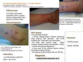
EM Clinical Case Presentation
EM Clinical Case Presentation. Arpan Patel. Triage. 25 yo Male with abdominal pain Afebrile , VSS - nausea + vomiting and diarrhea. History . 11 hours ago, sudden onset of sharp stabbing pain in the epigastric area (pt points there)

EM Clinical Case Presentation
E N D
Presentation Transcript
EM Clinical Case Presentation Arpan Patel
Triage • 25 yo Male with abdominal pain • Afebrile, VSS • - nausea • + vomiting and diarrhea
History • 11 hours ago, sudden onset of sharp stabbing pain in the epigastric area (pt points there) • Pain is non-radiating, not associated with meals or certain foods, and is not relieved by antacids. (He has been on Omeprazole for past 3 months) • Explains he has had similar episodes for the past 4 years, last was 2 weeks ago, pain abates when he takes 12 Tylenols at once. • Now denies nausea, vomiting, and diarrhea
More History • Denies chest pain, SOB, UTI sx, GERD sx, constipation, blood in stool, cough, and fever. • PMH: none • PSH: none • Meds: Omeprazole since 3/2012 • Allergies: NKDA • SH: Occasional smokes cigs, EtOH socially
Physical • BP 143/74, P 64, R 17, T 97.0F, O2 Sat: 100% on RA • General: Pt lying in bed, appears uncomfortable • CV: RRR, S1S2, no mrg • Resp: CTAB • Abd: soft, non-distended, tender in epigastrium and RUQ, no guarding/rebound, +bs in all quadrants • Back: No CVA tenderness
Differential Diagnosis • RUQ: - Cholelithiasis - Biliary Colic - Acute Cholecystitis - Cholangitis - Acute Hepatitis - Perf. Duodenal Ulcer - RLL Pneumonia • Epigastric: - Peptic Ulcer Disease - Pancreatitis - GERD - Myocardial Infarction • Atypical Px’s of: - Appendicitis - R sided Kidney disease: Pyelonephritis or Nephrolithiasis
Let’s get some Labs • BMP: 139/4.2 105/28.2 12/0.9 Glu 102 • CBC: 13.4 > 14.3/45.5 < 292 • UA completely negative • Coags, LFTs, Lipase and Amylase all wnl
For you EM US Fellowship Chasers: Bedside US: • Unable to visualize gallbladder, pt had not eaten in over 12 hours, but tells us he had an US done 4 years ago and was told he had many stones Official US: • Gallbladder completely filled with calculi limiting evaluation of GB wall thickness • Minimal pericholecystic fluid, no intra/extra hepatic bile duct dilation, CBD is 0.3 cm • Positive sonographic Murphy’s sign • R Kidney is 10.8 cm & nl, no hepatomegaly, some steatosis • Likely cholelithiasisvs Acute chole, can confirm with HIDA
Final Diagnosis • Acute Cholecystitis • Started on Cefoxitin, Morphine for pain, and admitted to surgery. • GB was removed 2 days later • Why treat/remove? Even though AC may resolve in 7-10 days on its own, it has a high rate of progressing to gangrenous chole and perforating which increases morbidity and mortality
Diagnostic Criteria for Acute Chole • Based on physical exam, labs, and imaging • Physical: RUQ pain, Murphy’s sign, fever, tachy • Labs: Leukocytosis (left shift), shouldn’t have elevated bili or AlkPhos in simple Acute Chole • Imaging: On US - GB wall thickening (>4-5 mm), pericholecystic fluid, sono Murphy’s sign. On HIDA - the GB will not be visualized due to obstruction of the cystic duct by a stone or edema
What about the AC triad of RUQ pain, fever, and leukocytosis? He was afebrile! • Retrospective review of pts with AC dx’d with HIDA: fever defined as oral T>100 and rectal T>104, and leukocytosis as 11k or more • Of these cases 52% were nongangrenous AC, 26% gangrenous AC, and 22% chronic chole • In those with non-gangrenous AC, 71% were afebrile, 32% lacked leukocytosis, 28% lacked both • In those with gangrenous AC, 59% afebrile, 27% no leukocytosis, 16% lacked both. • Take home point: Do not rely on the presence of fever and leukocytosis to make a diagnosis of AC! Gruber PJ, Silverman RA, Gottesfeld S, Flaster E. Presence of fever and leukocytosis in acute cholecystitis. Ann Emerg Med. 1996 Sep;28(3):273-7