Muscles and Structures of Forearm and Hand Extensor Compartment
240 likes | 273 Vues
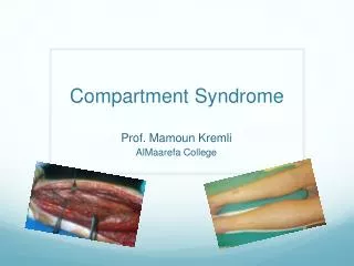
Explore the organization and function of muscles in the extensor compartment of the forearm and hand, along with the innervation, blood supply, and common injuries like tennis elbow and baseball fractures.

Muscles and Structures of Forearm and Hand Extensor Compartment
E N D
Presentation Transcript
Common Extensor Tendon Anconeus Posterior/Extensor fascial Compartment of fore arm Dorsum of the Hand
Objectives Fascial Compartmentalization Organization of Muscles according to their action Grouping of muscles into superficial & deep Vessels & nerve
Muscles Name them posterior extensor supinator All are innervated by radial nerve
Can be organized in threefunctional groups Muscles that extend abduct or adduct the hand at wrist extensor carpi radialis longus, extensor carpi radialis brevis & extensor carpi ulnaris Muscles that extend the medial four digits Extensor digitorum, extensor indices & extensor digiti minimi Muscles that extend or abduct thumb Abductor pollicis longus / brevis & extensor pollicis longus/brevis Anconeus Supinator & Brachioradialis
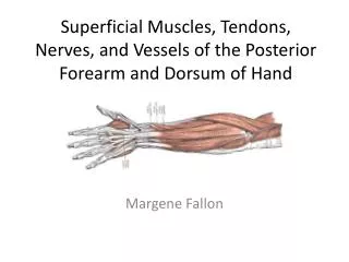
Posterior facial compartment Superficial group extensor carpi radialis brevis extensor digitorum extensor digiti minimi extensor carpi ulnaris anconeus Common Extensor Tendon Deep group Supinator, abductor pollicis longus extensor pollicis brevis extensor pollicis longus extensor indices Lateral facial compartment Brachioradialis Exten. Carpi radialis longus
Superficial group extensor carpi radialis brevis extensor digitorum, extensor digiti minimi, extensor carpi ulnaris anconeus Common Extensor Tendon Anconeus 5 1 4 2 3
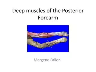
Deep group supinator abductor pollicis longus extensor pollicis longus extensor pollicis brevis extensor indices extensor indices
Lateral Facial Compartment Brachioradialis & extensor carpi radialis longus
Arteries of Posterior Compartment Anterior & posterior interosseous arteries arise from CIA a branch of ulnar artery
Nerves of Posterior Compartment Deep branch arises from radial nerve in front of the lateral epicondyle of humerus pierces the supinator & hinds around the neck of the radius, runs b/w the superficial & deep group. Gives muscular & articular branches
Structures pass superficial to the extensor retinaculum from medial to lateral Dorsal cutaneous br of ulnar nerve Basilic vein Cephalic vein Superficial br of radial nerve Structures pass deep to the extensor retinaculum from medial to lateral The tendons of: Ext carpi ulnaris Ext digiti minimi Ext digitorum Ext indicis Ext pollicis longus Ext carpi radialis longus Abductor pollicis longus Ext pollicis brevis
Extensor retinaculum Is a thickening of deep fascia that stretches across the back of the wrist & holds the long ext tendons. Attached medially to pisiform & hook of hamate & laterally to the distal end of radius.
Synovial tendon sheath 5 2 6 4 3 1
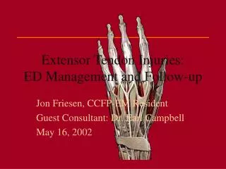
The Dorsum of the Hand Dorsal expansion 4 tendons of ED fan over the dorsum of the hand. Strong bands connect the tendons of little, ring & middle fingers proximal to the head of metacarpal bones. Each extensor tendon joins the extension expansion. Near the proximal interphalangeal joint extensor expansion split into 3 parts a central & 2 lateral parts, which converge to be inserted into the base of the distal phalanx
Tennis Elbow Tennis elbow is caused by the partial tearing or degeneration of the origin of the extensor muscles from lateral epicondyle of humerus Characteristics Tenderness over Pain radiating down the lateral side of the fore arm Common in tennis players, violinists & houswives