Download
1 / 15
180 likes | 588 Vues
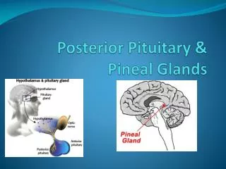
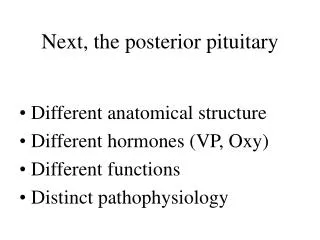
Next, the posterior pituitary. Different anatomical structure Different hormones (VP, Oxy) Different functions Distinct pathophysiology. Hypothalamic Control of Posterior Pituitary Secretion (a summary).

E N D
Next, the posterior pituitary • Different anatomical structure • Different hormones (VP, Oxy) • Different functions • Distinct pathophysiology
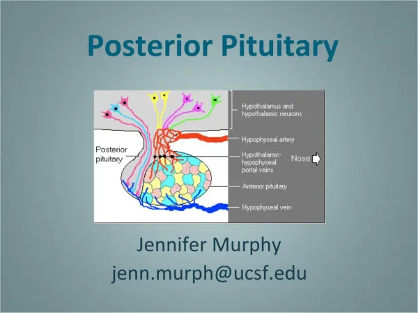
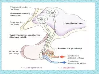
Hypothalamic Control of Posterior Pituitary Secretion (a summary) • Magnocellular neurons in SON and PVN synthesize precursor peptides for vasopressin (antidiuretic hormone) or oxytocin) • Products are packaged into neurosecretory vesicles and transported in axons forming the hypothalamo-hypophyseal tract • Vesicles are stored in posterior pituitary. • Release by exocytosis is controlled by neuroendocrine reflexes.
Neurohypophysial hormones:similar nonapeptides, derived from different precursors
Prepro-oxytocin -lys-arg- -lys-arg- H2N- -COOH H2N- Signal peptide Processing in rough ER (loss of signal peptide) H2N- H2N- Pro-oxytocin H2N- -lys-arg- -lys-arg- -COOH Processing in Golgi – hydrolysis of lys-arg bonds Oxytocin Neurophysin I Oxytosin carrier protein in axon Figure 2. Prepro-oxytocin. Proteolytic maturation proceeds from top to bottom
Prepro-vasopressin -lys-arg- -lys-arg- H2N- -COOH Signal peptide Processing in rough ER (loss of signal peptide) Pro-vasopressin H2N- -lys-arg- -lys-arg- -COOH Processing in Golgi – hydrolysis of lys-arg bonds Vasopressin Neurophysin II ADH carrier protein in axon Glycoprotein Figure 2. Processing of prepro-vasopressin.
Schema to represent steps in neuropeptide synthesis, transport and release
EM image of neurosecretory granules packed into Herring bodies in neurohypophysial axon terminals
Hormone storage and release from neurohypophysial axon terminals
Immunocytochemical visualization of vasopressin- and oxytocin-synthesizing neurons • Upper box, coronal section through the hypothalamic paraventricular nucleus (PVN) • Lower box, section through the hypothalamic supraoptic nucleus (SON) • VP, dark, Oxy light brown • NB: VP, Oxy in separate cells, applies to both male and female brain
Physiology of Oxytocin Secretion • In females, 2 unique roles: • Milk ejection: sensory stimulation of the nipple induces firing of oxytocin cells, release of oxytocin into the blood, activation of oxytocin receptors in breast myoepithelial cells and milk expulsion • Delivery of the fetus: distention of the uterus at term triggers firing of oxytocin neurons, releasing oxytocin as a hormone into the blood; occupany of oxytocin receptors in uterine smooth muscle induces contractions that assist in expulsion of the fetus.
Lactation is a cooperation between anterior and posterior pituitary hormones. Prolactin released from the anterior pituitary lactotrophs promotes milk production; oxytocin released from posterior pituitary storage sites promotes contraction of myoepithelial cells and milk expulsion
In-vivo electrophysiology of oxytocin-secreting neurosecretory neuronsprerequisite:antidromic identification to verify axon target
Rat hypothalamus exposed for in-vivo electrophysiologyUnder anesthesia, removal of the sphenoid bone and dura mater exposes the ventral surface of the hypothalamus from the optic chiasm to the posterior pituitary. A stimulating electrode positioned in the posterior pituitary allows activation of axon terminals of neurosecretory neurons. A recording micropipette positioned near the junction of the middle and anterior cerebral arteries serves to record extracellular activity from antidromically-identified supraoptic nucleus neurons.