Download
1 / 12
120 likes | 225 Vues
This review revealed a new mechanism for gene regulation through “gene silencing” at the transcriptional level (TGS) or at the posttranscriptional level (PTGS), which play a key role in many essential cellular processes. Today dsRNA is used as a powerful tool to experimentally elucidate the function of essentially any gene in a cell.

E N D
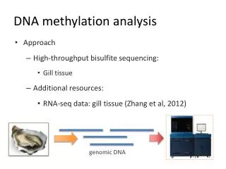
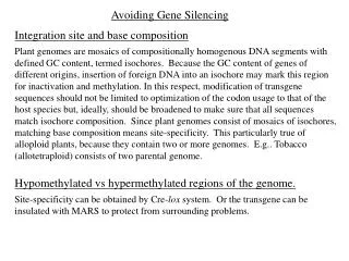
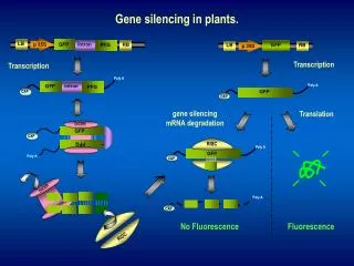
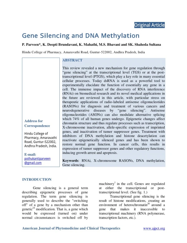
Original Article Gene Silencing and DNA Methylation P. Parveen*, K. Deepti Brundavani, K. Mahathi, M.S. Bhavani and SK. Shaheda Sultana Hindu College of Pharmacy, Amaravathi Road, Guntur-522002, Andhra Pradesh, India ABSTRACT This review revealed a new mechanism for gene regulation through “gene silencing” at the transcriptional level (TGS) or at the post- transcriptional level (PTGS), which play a key role in many essential cellular processes. Today dsRNA is used as a powerful tool to experimentally elucidate the function of essentially any gene in a cell. The immense impact of the discovery of RNA interference (RNAi) on biomedical research and its novel medical applications in the future are reviewed in this article, with particular stress on therapeutic applications of radio-labeled antisense oligonucleotides (RASONs) for diagnosis and treatment of various cancers and neurodegenerative diseases by oligonucleotides (ASONs) can also modulate alternative splicing which 74% of all human genes undergo. Epigenetic changes affect chromatin structure and thus regulate processes such as transcription, X-chromosome inactivation, allele-specific expression of imprinted genes, and inactivation of tumor suppressor genes. Treatment with inhibitors of DNA methylation and histone deacetylation can reactivate epigenetically silenced genes and has been shown to restore normal gene function. In cancer cells, this results in expression of tumor suppressor genes and other regulatory functions, inducing growth arrest and apoptosis. “gene silencing”. Antisense Address for Correspondence Hindu College of Pharmacy, Amaravathi Road, Guntur-522002, Andhra Pradesh, India. E-mail: pothukantiparveen @gmail.com Keywords: RNAi, X-chromosome RASONs, DNA methylation, Gene silencing. INTRODUCTION Gene silencing is a general term describing epigenetic processes of gene regulation. The term gene silencing is generally used to describe the "switching off" of a gene by a mechanism other than genetic24 modification. That is, a gene which would be expressed (turned on) under normal circumstances is switched off by machinery1 in the cell. Genes are regulated at either the transcriptional or post- transcriptional level. (See fig. 1.) Transcriptional gene silencing is the result of histone modifications, creating an environment of heterochromatin26 around a gene that makes transcriptional machinery (RNA polymerase, transcription factors, etc.). it inaccessible to American Journal of Phytomedicine and Clinical Therapeutics www.ajpct.org
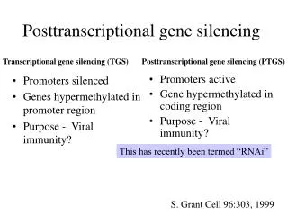
Parveen et al________________________________________________ ISSN 2321 –2748 Post-transcriptional gene silencing is the result of mRNA of a particular gene being destroyed. The destruction of the mRNA prevents translation to form an active gene product (in most cases, a protein). A common mechanism of post-transcriptional28 gene silencing is RNAi. Both transcriptional transcriptional gene silencing are used to regulate endogenous genes. Mechanisms of gene silencing also protect3 the organism's genome from transposons and viruses. Gene silencing thus may be part of an ancient immune system protecting from such infectious DNA elements. RNA interference The enzyme dicer trims double stranded RNA, to form small interfering RNA or micro RNA. These processed RNAs are incorporated into the RNA-induced silencing complex (RISC), which targets messenger RNA to prevent translation. RNA interference (RNAi) is a mechanism that inhibits gene expression by causing the degradation of specific RNA molecules or hindering the transcription of specific genes. The targets are often RNA from viruses and transposons14 (probably a form of innate immune response), although it also plays a role in regulating16 development and genome maintenance. Key to the RNAi processes are small interfering RNA strands (siRNA), which have nucleotide sequences to a targeted RNA strand. The siRNA "guides" proteins5 within the RNAi pathway to the targeted messenger RNA (mRNA) and "cleaves" them, breaking them down into smaller portions that can no longer be translated24 into protein. A type of RNA transcribed from the genome itself, miRNA, and works in the same way. The selective and robust effect of RNAi on gene expression makes it a valuable research tool, both in cell culture and in living organisms because introduced into cells can induce suppression of specific genes of interest. RNAi may also be used for large-scale systematically shut down each gene in the cell, which can help identify the components necessary for a particular cellular process7 or an event such as cell division. Exploitation of the pathway is also a promising tool in biotechnology6 and medicine. DNA methylation DNA methylation is a biochemical process that is important for normal development in higher organisms. It involves the addition of through cell a methyl group purine rin to the 5 position of the cytosine pyrimidine ring or the number 6 nitrogen of the adenineg (cytosine and adenine are two of the four bases of DNA). This modification can be inherited division. DNA methylation is a crucial part of normal organismal development2 and cellular differentiation in higher organisms. DNA methylation stably alters the gene expression pattern in cells such that cells can "remember where they have been" or decrease gene expression; for example, cells programmed to be pancreatic islets during embryonic development remain throughout the life of the organism without continuing signals telling them that they need to remain islets. DNA methylation is typically removed during zygote formation and re- established through successive cell divisions during development. However, the latest research shows that hydroxylation of methyl group occurs rather than complete removal of methyl groups in zygote. Some methylation modifications that regulate gene expression are inheritable and are referred to as epigenetic regulation. In addition, suppresses the expression of viral genes42 and other deleterious elements that have been incorporated into the genome of the host over time. DNA methylation also forms the basis screens that and post- pancreatic islets complementary DNA methylation synthetic dsRNA AJPCT[2][5][2014]544-555
Parveen et al________________________________________________ ISSN 2321 –2748 of chromatin structure, which enables cells to form the myriad characteristics necessary for multi cellular life from a single immutable sequence of DNA. Methylation also plays a crucial role in the development of nearly all types of cancer.DNA methylation at the 5 position of cytosine has the specific effacing gene expression and has been found in every vertebrate examined. In adult ct of redu somatic tissues, DNA methylation typically occurs in a CpG dinucleotide context; non- CpG methylation9 is prevalent in embryonic stem cells. Mechanism of DNA methylation In mammalian DNA, 5-methylcyto- sine is found in approximately 4% of genomic DNA, primarily at dinucleotides (CpGs). Such CpG sites occur at lower than expected frequencies throughout the human genome but are found more frequently at small stretches of DNA called CpG islands. These islands are typically found in or near promoter regions of genes, where transcription is initiated. In contrast to the bulk of genomic DNA, in which most CpG sites are heavily methylated, CpG islands in germ-line tissue and promoters of normal somatic cells remain un-methylated, allowing gene expression to occur. (See fig. 2.) DNA methylation helps to maintain transcriptional silence in non expressed or non coding regions of the genome. For example, pericentromeric11 heterochromatin, which is condensed and transcriptionally inactive, is heavily methylated. Hyper methylation thus ensures this DNA is late- replicating and transcriptionally quiescent, and suppresses the expression of any potentially harmful viral sequences or transposons that may have integrated into such sites containing highly repetitive sequences. By contrast, these sites are generally un-methylated in promoter regions of euchromatin, regardless transcriptional state of the gene. Exceptions to this rule, however, can be found in mammalian cells where these regions are methylated to maintain inactivation. Thus, CpG islands in promoters of genes located on the inactivated X chromosome of females are methylated, as are certain imprinted genes in which only the maternal or paternal allele is expressed. Epigenetic effects hypermethylation can also induce inevitable alterations in gene xpression. Methylation of the DNA repair genes MLH1 and MGMT can lead to their inactivation, methylation may affect the transcription of genes in two ways. First, the methylation of DNA itself may physically impede transcriptional proteins to the gene, and second, and likely methylated DNA may be bound by proteins known as methyl-CpG-binding domain proteins (MBDs). MBD proteins then recruit additional proteins to the locus, such as histone deacetylases and other chromatin remodeling proteins that can modify histones, thereby forming compact, inactive chromatin, termed heterochromatin. This link between DNA methylation and chromatin structure is very important. In particular, loss of methyl- CpG-binding protein 2 (MeCP2) has been implicated in Rett medimethyl-CpG-binding domain protein 2 (MBD2) ates the transcriptional silencing of hypermethylated genes in cancer. Research has suggested that long-term memory storage in humans may be regulated by DNA methylation. Regulation of DNA methylation DNA methylation is controlled at several different levels in normal and tumor cells. The addition of methyl groups is carried out by a family of enzymes, DNA methyltransferases (DNMTs). structure in the vicinity of gene promoters also affects DNA transcriptional such as the binding of cytosine-guanosine more important, syndrome; and Chromatin of the methylation and AJPCT[2][5][2014]544-555
Parveen et al________________________________________________ ISSN 2321 –2748 transcriptional activity. These are in turn regulated by various factors such as nucleosome spacing and histone acetylases13, which affect access to transcriptional factors. DNA methyltransferases In mammalian methylation occurs mainly at the C5 position of CpG dinucleotides and is carried out by of enzymatic activities–maintenance methylation and de novo methylation. Two general classes. Maintenance methylation activity is necessary to preserve DNA methylation after every cellular DNA replication cycle. Without the DNA methyltransferase (DNMT), the replication machinery itself would produce daughter strands that are un-methylated and, over time, would demethylation. DNMT1 is the proposed maintenance methyltransferase responsible for copying DNA methylation15 patterns to the daughter strands during DNA replication. Mouse models with both copies of DNMT1 deleted are embryonic lethal at approximately day 9, due to the requirement of DNMT1 activity for development in mammalian cells. It is thought that DNMT3a and DNMT3b are the de novo methyltransferases that set up DNA methylation patterns early in development. DNMT3L is a protein that is homologous to the other DNMT3s but has no catalytic activity. Instead, DNMT3L assists the de novo methyltransferases by increasing their ability to bind to DNA and stimulating their activity. Finally, DNMT2 (TRDMT1) has been identified as a DNA methyl- transferase homolog, containing all 10 sequence motifs common to all DNA methyltransferases; (TRDMT1) does not methylate DNA but instead methylates anticodon loop of aspartic acid transfer RNA. Histone deacetylation Although methylation controls gene activity, it alone is insufficient to repress transcription. The local chromatin structure also contributes in determining whether genes are transcribed or repressed. For example, DNMTs may regulate gene accompanying promoter DNA methylation by recruiting histone deacetylases (HDACs) and other chromatin-binding proteins to promoter sites to maintain histone deacetylation. The state of histone acetylation is important in regulating chromatin17 structure and gene transcription. The chromatin structure surrounding transcriptionally active genes differs from that of methylated, silenced nucleosome structure and histone acetylation affect chromatin structure and thus regulate gene transcription. composed of 146 base pairs of DNA wound around complexes of eight histones within the nucleus. In CpG islands that are un- methylated, the nucleosomes are in a more open configuration that allows access of factors that favor transcription When CpG islands are hypermethylated, heterochromatin, the nucleosomes become more tightly compacted, inhibiting the access of regulatory proteins transcription. Interestingly, recent studies have determined that methylation of specific residues on the tails of histone H3 are critical for maintaining the appropriate chromatin configuration. Methylation of lysine 9 residues on H3 facilitates transcriptional repression, whereas methylation of lysine 4 on H3 is associated with transcriptionally active euchromatin. (See fig. 3.) Histone deacetylation inhibitors Given that HDACs are integral to maintaining transcriptional silence, they can increase gene expression in the absence of hypermethylation19. A variety of HDAC inhibitors have been shown to inhibit histone cells, DNA silencing un-methylated, genes. Both lead to passive Nucleosomes are that is as in that promote however, DNMT2 cytosine-38 in the AJPCT[2][5][2014]544-555
Parveen et al________________________________________________ ISSN 2321 –2748 deacetylation in human tumor cells, leading to accumulation of acetylated histone proteins. In leukemic cells, treatment with HDAC inhibitors results differentiation, or apoptosis. These drugs hold promise for cancer therapy, especially when used in combination with methylation inhibitors, other differentiating agents, or cytotoxic compounds. HDAC inhibitors undergoing clinical evaluation for treatment of hematopoietic malignancies include the natural HDAC inhibitor hydroxamic acid derivatives (such suberanilohydroxamic tetrapeptide depsipeptide50, derivatives like MS-275 and CI-994, and aliphatic acids such as valproic acid and phenylbutyrate. It should be noted that methylation is dominant to histone deacetylation, so transcription cannot occur without first inhibiting methylation. Initial treatment with azacitidine or decitabine, followed by HDAC inhibitors, can produce additive or synergistic effects for re-expression of transcriptionally silenced genes These preclinical findings, coupled21 with the above model of chromatin structure and its regulation methylation and acetylation, provide the rationale for therapeutic approaches in which the two processes are targeted22. Gene methylation in cancer According to the widely accepted 'two-hit' hypothesis proposed by Knudson, loss of function of both alleles in a given gene (e.g. a tumor suppressor) is required transformation. The first hit typically is in the form of a mutation in a critical gene, followed by loss of the wild-type allele through deletion32 or loss of heterozygosity. In familial cancers this first hit may occur through germ-line mutations, while in noninherited sporadic mutations are more commonly observed. Subsequent loss of the remaining allele through deletion, point mutation, or loss of heterozygosity can eliminate the remaining functional gene. Inactivation of regulatory genes in this manner can lead to the development of cancer. DNA methylation is an important regulator of gene transcription and a large body of evidence has demonstrated that aberrant DNA methylation is associated with unscheduled gene silencing, and the genes with high levels of 5-methylcytosine23 in their promoter region are transcriptionally silent. DNA methylation49 embryonic development, and in somatic cells, patterns of DNA methylation are generally transmitted to daughter cells with a high fidelity. Aberrant DNA methylation patterns have been associated with a large number of human malignancies and found in two distinct forms: hypermethylation and hypomethyla- tion compared to Hypermethylation is one of the major epigenetic25 modifications transcription via promoter region of tumour suppressor genes. Hypermethylation typically occurs at CpG islands in the promoter region and is associated with gene inactivation. Global hypomethylation has also been implicated in the progression27 of cancer through different mechanisms. (See fig. 4.) Demethylation and gene reactivation The demethylating agent azacitidine and its deoxy derivative, decitabine, are powerful inhibitors of DNA methylation. Preclinical studies have shown that after cellular uptake and azacitidine is incorporated into RNA, where it suppresses RNA synthesis and has cytotoxic effects. After conversion deoxycytidine diphosphate47 by ribonucle- otide reductase phosphorylation, the triphosphate form is in growth arrest, trichostatin, as acid), the benzamide cyclic is essential during normal tissue. that repress by both simultaneously development and of carcinogenesis for malignant phosphorylation, to 5-aza-2'- and subsequent cancers somatic AJPCT[2][5][2014]544-555
Parveen et al________________________________________________ ISSN 2321 –2748 incorporated into DNA in place of the natural base cytosine. Because of the substitution of the 5' nitrogen atom in place of the carbon, the DNMTs are trapped29 on the substituted DNA strand and methylation is inhibited. Thus, in the presence of these analogs, a significant proportion of the DNA becomes hemimethylated41. A second round of DNA synthesis in the presence of these drugs results in full double-stranded demethylation while both azacitidine and decitabine inhibit methylation, they do differ in some respects. Because it contains a ribose moiety, azacitidine can be phosphorylated by uridine-cytidine kinase, and when this happens it is subsequently incorporated into RNA. Since decitabine deoxyribose31 group, it is incorporated only into DNA. While some have suggested that differential incorporation into RNA and DNA may account for differences in methylation and toxicity profiles between these two agents,there are no direct clinical data at present to support this proposition. Inhibition of Methylation with azacytidine See figure 5. Detectors DNA methylation can be detected by the following assays currently used in scientific research: Methylation-Specific PCR (MSP), which is based on a chemical reaction of sodium bisulfite with DNA39 that converts un- methylated cytosines dinucleotides to uracil or UpG, followed by traditional PCR. However, methylated cytosines will not be converted in this process, and primers are designed to overlap the CpG site of interest, which allows one to determine methylation status as methylated or un-methylated. Whole genome bisulfite sequencing, also known as BS-Seq, which is a high- throughput genome-wide analysis of DNA methylation. It is based on aforementioned sodium conversion of genomic DNA, which is then sequencing on a Next-generation sequencing platform. The sequences obtained are then re-aligned to the reference genome methylation states of CpG dinucleotides based on mismatches resulting from the conversion of unmethylated cytosines into uracil. The HELP assay, which is based on restriction enzymes' differential ability to recognize and cleave methylated and unmethylated CpG DNA sites. Now rarely-used assay based upon restriction enzymes' differential recogni- tion of methylated and unmet ChIP-on- chip assays, which is based on the ability of commercially prepared antibodies to bind to DNA methylation-associated proteins33 like MeCP2. Restriction landmark genomic scanning, a complicated and ylated CpG sites; the assay is similar in concept to the HELP assay. Methylated DNA immunoprecipitation (MeDIP), analogous immunoprecipitation, tation is used isolate methylated DNA fragments for input into DNA detection methods such as to DNA microarrays (MeDIP-chip) or (MeDIP-seq). Future challenges The age in which epigenetics becomes an essential component in the clinical management of the clinical management of the oncology patient is getting closer Treatment of age macular degeneration Prevents brain damage caused by huntingtons disease Treatment of alzimers disease and spinal cord injury bisulfite to determine DNA contains a to immunoprecipi- chromatin DNA sequencing of CpG AJPCT[2][5][2014]544-555
Parveen et al________________________________________________ ISSN 2321 –2748 4. Treatment of developmental abnormali- ties. CONCLUSION The biological significance of DNA methylation in the regulation of gene expression and its role in cancer is increasingly recognized. It is now clear that these epigenetic changes affect chromatin structure and thus regulate processes such as transcription, X-chromosome inactivation, allele-specific expression of imprinted genes, and inactivation of tumor suppressor genes. Treatment with inhibitors methylation and histone deacetylation can reactivate epigenetically silenced genes and has been shown to restore normal gene function. In cancer cells, this results in expression of tumor suppressor genes and other regulatory functions, inducing growth arrest and apoptosis. decitabine are clinically active in MDS and other hematologic malignancies and may have therapeutic potential in other malignant disorders. Continued clinical trials with hypomethylating agents inhibitors, alone or in combination, may provide further advances in the treatment of hematopoietic malignancies and solid tumors. Epigenetics is everywhere you can inherit something beyond DNA sequence that’s were the real excitement in genetics now. REFERENCES 1. Bertil D aneholt. T he Nobel Prize in physiology and Medicine 2006. The Nobel Assembly at Karolinska Institute. 2. Fire A, XuS, Montgomery MK, Kostas SA, D river SE and Mello- CC: Potent and specific genetic inter ference by double stranded RNA in aenorhabditis elegans. Nature 1998; 391:806-11. 3. Hannon GL. RNA interference. Nature 2002; 418:244-51. Mello CC, C onte D. Revealing the world of RNA interference Nature 2004; 431:338- 342. Meuster G, T uschl T . Mechanisms of gene silencing by double stranded RNA. Nature 2004; 431:343-9. Hammond SM. D icing and Slicing. T he core machinery of the RNA inter ference pathway. FE BS Letters 2005; 579: 5822-9. Plastre K. RHA RNA silencing: the Genome’s immune system. Science 2002; 296:1263-65. Dorsett Y, T uschl T. SiRNA- applications in functional genomics and potentials in therapeutic. Nature Reviews 2004. Hannon GJ, Rose JJ. Unlocking the potential of the human genome interference. Nature 2004; 431:371-8. 10.Soutschek J, et al. T herapeutic silencing of an endogenomic administration of modified SiRNA. Nature 2004, 432, 173-178. 11.Morrsey D V, et al. Potent and persistent invivo antiHBV activity of chemically modified siRNA. Nature 2005;23: 1002-7. 12.Simmerman T S, et al. RNAi mediated gene silencing in nonhuman primates. Nature 2006; 441:111-4. 13.Deewanjee Mrinal K, et al. D evelopment of sensitive radioiodinated oligonucleotide probes techniques. Bioconjug C hem 1992; 2:195- 200. 14.Hnatowich D J. Antisense imaging. Where are we now? Cancer Biother Radiopharm 2000; 15:447-57. 15.Askari F K, Mc D onnell WM. Antisense oligonuclotide therapy. 334:316-8. 16.Wickstrom E. C linical trials of genetic therapy with antisense D NA and D NA vectors. Ibid- ASON for cMYC mRNA in Burkitt’s lymphoma. 17.Nature clinical practice oncology dna methylation and gene silencing in cancer. 18.The epigenetic progenitor origin of human cancer. Andrew P. Feinberg Nature Reviews, volume. 19.Epigenetics and cancer treatment. european journal of pharmacology. 5. 6. 7. 8. 9. of DNA with RNA gene by systemic Azacitidine and and HDAC antisense conjugation by NEJM 1996; AJPCT[2][5][2014]544-555
Parveen et al________________________________________________ ISSN 2321 –2748 20.The cotext and potential of epigenetics in oncology. J Lopez, M perchade, HM coley , A Webb and T crook. 21.McLaughlin, C. K.; Sleiman, H. F. assembly Chem. Soc. Rev. 2011, 40, 5647– 56. 22.Fu, J.; Liu, M.; Liu, Y.; Yan, H. Spatially- interactive biomolecular networks organized by nucleic acid nanostructures Acc. Chem. Res. 2012, 45, 1215–26. 23.Campolongo, M. J.; Tan, S. J.; Xu, J.; Luo, D.DNA nanomedicine: Engineering DNA as a polymer for therapeutic and diagnostic applications Adv. Drug Delivery Rev. 2010, 62,606–616. 24.Smith, D.; Schuller, V.; Engst, C.; Radler, J.; Liedl, T. Nucleic acid nanostructures for biomedical cine. 2013, 8, 105-21. 25.Hamblin, G. D.; Fakhoury, J. F.; Bujold, K. E.; Sleiman, H. F. Rolling circle amplification-templated DNA nanotubes show increased stability and cell penetration ability J. Soc. 2012, 134, 2888–91. 26.Walsh, A. S.; Yin, H.; Erben, C. M.; Wood, M. J. A.; Turberfield, A. J.DNA cage delivery to mammalian Nano 2011, 5, 5427–5432. 27.Lee, H.; Lytton-Jean, A. K. R.; Chen, Y.; Love, K. T.; Park, A. I.; Karagiannis, E. D.; Sehgal,A.; Querbes, W.; Zurenko, C. S.; Jayaraman, M.; Peng, C. G.; Charisse, K.; Borodovsky, A.; Donahoe, J. S.; Truelove, J.; Nahrendorf, M.; Langer, R.; Anderson, D. G. Molecularly self-assembled nucleic acid nanoparticles for targeted in vivo siRNA delivery Nat. Nanotechnol. 2012, 7, 389–393. 28.Keum, J.-W.; Ahn, J.-H.; Design, assembly, and activity of antisense DNA nanostructures Small 2011, 7, 3529– 3535. 29.Schuller, V. J.; Heidegger, S.; Sandholzer, N.; Nickels, P. C.; Endres, S.; Bourquin, C.; Liedl, T. Cellular immunostimulation coated DNA origami Nano 2011, 5, 9696–9702. 30.Li, J.; Pei, H.; Zhu, B.; Liang, L.; Wei, M.; He, Y.; Chen, N.; Li, D.; Huang, Q.; Fan, C. Self-assembled nanostructures for noninvasive intracellular delivery of immunostimulatory oligonucleotides ACS Nano 2011, 5, 8783– 8789. 31.Jiang, Q.; Song, C.; Nangreave, J.; Liu, X.; Lin, L.; Qiu, D.; Wang, Z.-G.; Zou, G.; Liang, X.; Yan, H.; Ding, B.DNA origami as a carrier for circumvention of drug resistance J. Am. Chem. Soc. 2012, 134, 13396–13403. 32.Kim, K. R.; Kim, D. R.; Lee, T.; Yhee, J. Y.; Kim, B. S.; Kwon, I. C.; Ahn, D. R. Drug delivery by a tetrahedron for overcoming drug resistance in breast cancer cells Chem Comm. 2013, 49, 2010–2. 33.Modi, S.; M, G. S.; Goswami, D.; Gupta, G. D.; Mayor, S.; nanomachine that maps spatial and temporal pH changes inside Nanotechnol. 2009, 4,325–30. 34.Conway, J. W.; Castor, K. J.; Sleiman, H.DNA nanostructure serum stability: Greater than the sum of its parts Chem. Commun. 2013, 49, 1172–4. 35.Edwardson, T. G. W.; Carneiro, K. M. M.; McLaughlin, C. K.; Sleiman, H. F. Site-specific positioning of dendritic alkyl chains on DNA cages enables their geometry-dependent self-assembly Nat. Chem. 2013, 5, 868–875. 36.Lo, P. K.; Karam, P.; McLaughlin, C. K.; Cosa, G.; Sleiman, H. F. Loading and selective release of cargo in DNA nanotubes with longitudinal variation Nat. Chem. 2010, 2, 319–28. 37.Keum, J.-W.; Bermudez, H. resistance of DNA nanostructures to enzymatic digestion Chem. 2009, 7036–7038. 38.Davidson, B. L.; McCray, P. B., Jr.Current prospects for RNA therapies Nat. Rev.: Genet. 2011, 12, 329– 40. 39.DeVos, S. L.; Miller, T. M. Antisense oligonucleotides: multivalent DNA Hamblin, G. Supramolecular D.; DNA CpG self-assembled DNA applications Nanomedi- Carneiro, K. M.; Krishnan, Y.A DNA living cells Nat. McLaughlin, C. K.; Am. Chem. cells ACS Serpell, C. J.; Manoharan, M.; Aldaye, F. Hamblin, G. A.; D.; Bermudez, H. Enhanced Commun. Suhartha, N. A.; interference-based by CpG-sequence- structures ACS Treating neuro- AJPCT[2][5][2014]544-555
Parveen et al________________________________________________ ISSN 2321 –2748 degeneration RNA Neurotherapeutics 2013, 10, 486–97. 40.Deleavey, G. F.; Watts, J. K.; Alain, T.; Robert, F.; Kalota, A.; Pelletier, J.; Gewirtz, A. M.; Sonenberg, N.; Damha, M. J. Synergistic effects between analogs of DNA and RNA improve the potency of siRNA-mediated silencing Nucleic Acids Res. 2010, 38, 4547– 4557. 41.Oliveira, C. L.; Juul, S.; Jorgensen, H. L.; Knudsen, B.; Tordrup, D.; Falconi, M.; Koch, J.; Pedersen, J. S.; Andersen, F. F.; Knudsen, B. R. Structure of octahedral DNA cages: Variation of single- stranded linker regions and influence on assembly yields ACS Nano 2010, 4, 1367– 76. 42.Ferrari, N.; Bergeron, D.; Tedeschi, A. L.; Mangos, M. M.; Paquet, L.; Renzi, P. M.; Damha, M. J. Characterization of antisense oligonucleotides comprising 2′-deoxy-2′- fluoro-beta-d-arabinonucleic acid (FANA): Specificity, potency, and duration of activity Ann. N.Y. Acad. Sci. 2006, 1082, 91– 102. 43.Kanaori, K.; Tamura, Y.; Nishi, M.; Kanehara, H.; Tajima, K.; Makino, K. Structure and stability of the consecutive stereoregulated chiral phosphorothioate duplex Biochemistry 1999, 38, 160. 44.Astriab-Fisher, A.; Juliano, R.; Herdewijn, P. Increased uptake at the level of of antisense oligonucleotides by delivery as double stranded Pharmacol. 2004, 68(3) 403–7. 45.Bielinska, A.; Shivdasani, R. A.; Zhang, L. Q.; Nabel, G. J. Regulation of gene expression with phosphorothioate Science 1990, 250, 997–1000. 46.Kim, S. H.; Jeong, J. H.; Lee, S. H.; Kim, S. W.; Park, T. G.PEG conjugated VEGF siRNA for anti-angiogenic gene therapy J. Controlled Release 2006, 116, 123–9. 47.Breunig, M.; Hozsa, C.; Watanabe, K.; Umeda, I.; Goepferich, A. Mechanistic investigation of poly (ethylene imine)-based siRNA delivery: Disulfide bonds boost intracellular release of the cargo J. Controlled 130, 57–63. 48.Josse J, Kaiser Ad, kornberg A. Enzymatic synthesis of deoxyribonucleic acid. VIII. Frequencies of nearest neighbor base sequences in deoxyribonucleic acid. J Biol Chem. 1961 Mar; 236:864–875. [PubMed] 49.Swartz Mn, Trautner Ta, Kornberg A. Enzymatic synthesis of deoxyribonucleic acid. XI. Further studies on nearest neighbor base sequences in deoxyribonucleic acids. J Biol Chem. 1962 1967. [PubMed]. 50.Bird AP, Taggart MH. Variable patterns of total DNA and rDNA methylation in animals. Nucleic Acids Res. 1980 Apr 11; 8(7):1485–1497. [PMC [PubMed]. complexes Biochem. Aishwarya, V.; double-stranded oligonucleotides gene Oteri, F.; Desideri, A.; Lungwitz, U.; Kato, H.; nanoscale truncated Release 2008, Wada, T.; Morii, T.; Jun; 237:1961– DNA Fisher, M. H.; free article] AJPCT[2][5][2014]544-555
Parveen et al________________________________________________ ISSN 2321 –2748 Figure 1. Transcriptional RNA polymerase Figure 2. HDC attaches via meCP AJPCT[2][5][2014]544-555
Parveen et al________________________________________________ ISSN 2321 –2748 Figure 3. The mechanism whereby DNA methylation and histone deacetylation cooperate to repress transcription AJPCT[2][5][2014]544-555
Parveen et al________________________________________________ ISSN 2321 –2748 Figure 4. Altered DNA-methylation patterns in tumorigenesis Figure 5. Pathway for the methylation of cytosine in the mammalian genome and effect of inhibiting methylation with 5- azacytidine AJPCT[2][5][2014]544-555