Download
1 / 9
90 likes | 117 Vues
The phytochemical properties and antimicrobial activities of extracts of Moringa oleifera whole seeds and dehusked seeds with methanol, ethyl acetate and water have been studied. The result of the phytochemical evaluation and thin layer Chromatogram showed that the extracts of the whole seeds compose of different secondary metabolites from the dehusked seeds.

E N D
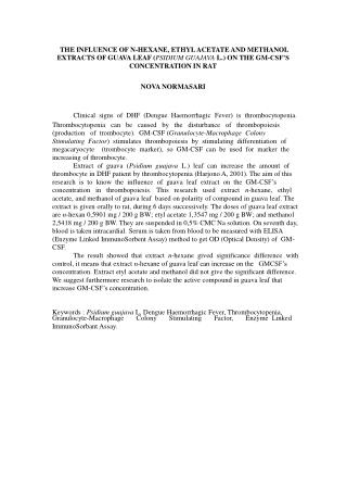
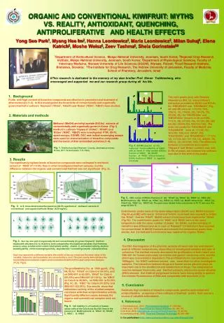
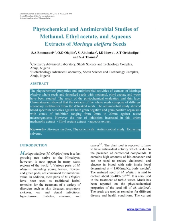
American Journal of Ethnomedicine, 2014, Vol. 1, No. 5, 346-354 Available online at http://www.ajethno.com © American Journal of Ethnomedicine Phytochemical and Antimicrobial Studies of Methanol, Ethyl acetate, and Aqueous Extracts of Moringa oleifera Seeds S.A Emmanuel*1, O.O Olajide1, S. Abubakar2, I.D Idowu1, A.T Orishadipe1 and S.A Thomas1 1Chemistry Advanced Laboratory, Sheda Science and Technology Complex, Abuja, Nigeria 2Biotechnology Advanced Laboratory, Sheda Science and Technology Complex, Abuja, Nigeria ABSTRACT The phytochemical properties and antimicrobial activities of extracts of Moringa oleifera whole seeds and dehusked seeds with methanol, ethyl acetate and water have been studied. The result of the phytochemical evaluation and thin layer Chromatogram showed that the extracts of the whole seeds compose of different secondary metabolites from the dehusked seeds. The antimicrobial study showed broad spectrum activities against both gram negative and gram positive organisms with zones of inhibition ranging from 9mm to 20mm against tested microorganisms. However the rate of inhibition increased in this order: methanolic extract > Ethyl acetate extract > aqueous extract. Keywords-Moringa oleifera, Phytochemicals, Antimicrobial study, Extracting solvents. cancer1-3. The plant pod is reported to have to have antioxidant activity which is due to the presence of carotenoid compounds. It contains high amounts of bio-enhancer and can be used to reduce cholesterol and glucose in blood with safe intake level determined at < 1.000mg/Kg body weight4. The matured seed of M. oleifera is said to contain about 38-40% oil3,2,5. It is also used in the treatment of turbid water. Much has been reported on the physicochemical properties of the seed oil of M.oleifera3. The seeds are used as remedies for different disease and health conditions. The current INTRODUCTION Moringaoleifera (M. Oleifera) tree is a fast growing tree native to the Himalayas, however, is now grown in many warm regions of the world1,2. Various parts of M. oleifera, including young leaves, flowers, and green pods, are consumed for nutritional value. In addition, most parts of M. Oleifera have been used as traditional herbal remedies for the treatment of a variety of disorders such as skin diseases, respiratory sickness, ear and hypertension, diabetes, dental infections, anaemia, and www.ajethno.com
American Journal of Ethnomedicine ________________________________________ ISSN: 2348-9502 work is aimed at studying the phytochemical and antimicrobial effect of the extracts of M. oleifera seeds using different solvents in relation to its application in folkloric medicine. MATERIALS AND METHODS anthraquinones, saponins, fats and oil6,7. Phenols Equal volumes of each extract and ferric chloride solution (which is prepared by dissolving 135.2g of FeCl3.6H2O in distilled water containing 20 ml of concentrated HCl dilute to 1 liter) are added together. A deep bluish green precipitate indicates the presence of phenol. Alkaloids To each extract was added to 1% aqueous HCl over water bath and filtered. The filtrate was treated with (2g of Iodine in 6g of Potassium iodide in 100 ml of distilled water). Formation brown or reddish brown precipitate indicates presence of alkaloids. Steroids Into each extract was added to 2ml acetic anhydride and 2ml H2SO4. Color change from violet to blue or green indicates the presence of steroids. Terpenes To each extract was added to 0.5ml acetic anhydride and a few drops of concentrated H2SO4. precipitate indicates terpenes. Cardiac glycosides The extract was treated with 2ml glacial acetic acid with a drop of Ferric Chloride solution and underplayed with 1ml H2SO4. A browning at the interface indicates the presence of cardiac glycosides. Tannins Each Extract was boiled in 20ml water and filtered. A few drops of 0.1% Ferric Chloride solution were added. Brownish green or indicates the presence of Tannins. cardiac glycosides, The plant was authenticated by Professor O. Olorode Department of Biological Science, Botany Unit, University of Abuja, Nigeria. Matured seeds of the Moringa oleifera plant were harvested from the Chemistry Advanced Moringa tree plantation and were identified by their physical properties as described by Price 200714. They were removed from the pod and dried further. The seeds were prepared as both whole seeds and dehusked seed. The whole seeds were first milled into powder with the aid of an electrical grinder and stored in moisture – free container, then the dehusked seeds were also milled into powder and stored comparison. Extraction of the plant sample About 300 ml of distilled methanol, ethyl acetate and water each of the solvent was measured and added to the 100g of the blended mixture of husk and seeds first, also with seed only in a stoppered glass container. The mixture was left for three days for extracting and then finally filtered. The methanol, ethyl acetate and aqueous extracts for dehusked seeds and whole seed were concentrated on the water bath and stored for subsequent analysis. Phytochemical screening For the purpose of this study, phytochemical screenings were carried out on the extracts to confirm the presence or absence of the following plant secondary metabolites: alkaloids, phenols, sterols, terpenes, tannins, Laboratory separately for A the bluish presence green of blue-black color flavonoids, Page 346-354
American Journal of Ethnomedicine ________________________________________ ISSN: 2348-9502 Flavonoids 5ml Ammonium solution was added to the aqueous filtrate of each extract and then a few drops of concentrated H2SO4. Yellow coloration indicates the presence of Flavonoids. Anthraquinones 10ml benzene was added to each extract and filtered. 0.5ml of 1% Ammonium solution was added and shaken. Pink, red, or violet color in the ammoniacal lower phase indicates the presence of Anthraquinones. Saponins 1g each extract was boiled with 5ml distilled water and filtered. 3ml distilled water was added to the filtrate and shaken vigorously for 5 minutes. Persistent frothing on warming indicates the presence of Saponins. Fats and oils Small quantity of each extract was pressed between two filter papers. Oily stains indicate the presence of Fats and s Oils. Triterpenoids Crude extract was mixed with chloroform and few drops of conc. H2SO4 was added, shaken and allowed to stand for some time. The formation of a yellow colored layer indicates the presence of triterpenoids. Glycosides 5ml H2SO4 was added to each of the test extract in a boiling tube. The mixture was heated in boiling water for 15minutes. Fehling’s solution A and B was added and the resulting mixture was heated to boiling. A brick red precipitates indicate the presence of glycosides. Carbohydrates 5ml of the equal mixture of both Fehling’s solutions A and B was added to 2ml of test extract in a boiling tube, this was heated for 2 minutes. A brick red precipitates indicate a positive result. Phlobatannins A few drops of 1% HCl were added to 1ml of test extract and was boiled. A reddish precipitates indicate the presence of phlobatannins. Resins 2ml of test extract plus an equal volume of acetic anhydride solution with drops of conc. H2SO4 gives a colophony resin, a violet color indicates the presence of resins. Balsams 3 drops of alcoholic FeCl3 were added to 4ml of extract which was warmed. A dark green coloration indicates the presence of the balsams. Volatile oil A small quantity of the test extract was shaken with dilute NaOH and 0.1M HCl. The formation of a white precipitate indicates a positive result. Antibacterial assays The antibacterial assay was carried out using agar well diffusion tests. The antimicrobial activity of the plant extracts was tested on five bacterial clinical isolates namely; Escherichia pneumonia, Proteus mirabilis, Pseudomonas aeruginosa and Staphylococcus aureus, the first four species represent gram negative bacteria while the last represent gram positive bacteria, these bacteria are the primary causative agents responsible for various human diseases conditions namely; urinary tract infections, arthritis and bone infections, diarrheal diseases, respiratory coli, Klebsiella Page 346-354
American Journal of Ethnomedicine ________________________________________ ISSN: 2348-9502 infections and meningitis among others. The test organisms were gotten from patients attending Specialist hospital Gwagwalada, F.C.T. Abuja and were further isolated and characterized using various biochemical tests adopted from8. Inoculum standardisation The microorganisms were inoculated into Mueller Hinton broth and incubated at 35 ± 2°C for 24 h. The turbidity of the suspensions scanned at 600 nm with an Ultra Violet Visible Spectrophotometer (Cecil, C-7500, Cambridge, England) the absorbance’s were measured and diluted with normal saline to compare with 1 McFarland turbidity standard. This level of turbidity is equivalent to approximately 3.0 × 10⁸ CFU/ml.9 Agar well diffusion assay The agar well diffusion technique was the standard method used to determine the antibacterial activity of the bioactive compounds. The culture medium (Mueller Hinton agar) was prepared and treated according to the manufacture's instructions. The agar plates were incubated for 24 h at 37°C to confirm their sterility. Absence of growth after 24 h showed that the plates were sterile. The sterile Mueller Hinton agar plates were inoculated with the test culture by surface spreading using a sterile cotton board and each bacterium evenly spread on the entire surface of the plate to obtain uniformity of the inoculum. The culture plate, then had five wells of 8 mm diameter made into it using a sterile cork borer. Streptomycin was used as a positive control at a concentration of (25µg/ml), while DMSO and distilled water (50:50v/v) was used as a negative control. Approximately, 0.2ml of the crude bioactive test compound of the various concentrations achieved using serial dilutions (50, 25, 12.5 µg/ml) were suspended in each wells and thereafter inoculated plates were incubated for 24 h at 37°C.The plates were examined for the presence of bacterial inhibition zones around each well. Antibacterial activities were determined by the presence of a zone of inhibition around the wells. The zones of inhibition were measured using a centimeter ruler and converted to the nearest millimeter (mm)16. RESULTS The aqueous and methanol extracts of the samples gave brown sticky solids after drying while the ethyl acetate extract was purely oil. This indicates that the solvents extracted different components from the seeds as revealed in the phytochemical result in Table 1. The result indicates the presence of more secondary metabolites in the ethyl acetate and methanol fractions while the aqueous fraction of the whole seeds tested negative to most of the constituents analyzed. Tannins, phenols, phlobatannins and resins tested negative in all the extracts. The results of the antimicrobial assay reveal that the antibacterial properties in various degrees on test organisms. From Table 2 below, the dehusked seed methanolic extract shows great actions on all the gram negative bacteria tested. The highest zones of inhibitions were pronounced on Proteus mirabilis (20.0±00mm) at 50µg/ml while the zone of inhibition of the remaining three gram negative organisms ranged between (18.0±00 to 18.5±00mm). The extract also shows activity on (14.5±00mm) at 50µg/ml which qualified it as a broad spectrum extract. The methanol extract of whole seeds shows activities on all the bacterial tested with the highest zone of (17.5±0.5mm) on Proteus mirabilis at concentration of 50µg/ml, the extract also possessed activity on Staph aureus at all the various working concentrations. The ethyl carbohydrates, extracts possessed Staph. Aureus Page 346-354
American Journal of Ethnomedicine ________________________________________ ISSN: 2348-9502 acetate extracts of the dehusked and whole seeds on Table 4 and 5 showed that the activity of the extract range between (15.0±00 to 9.0±00mm) which indicated a weak activity when compared with that of methanol extracts on each test microorganisms. The aqueous extract of the dehusked seeds shows activities on only two test organisms Proteus mirabilis and Klebsiella pneumonia (9.0±00mm) at 50µg/ml each respectively. The result of the aqueous whole seed extract showed activities on E. coli and Proteus mirabilis in all the working concentration while no activities were recorded on Staph. aureus. DISCUSSION could serve as potent starting material in the synthesis of these hormones. The present result also suggests that the matured seeds may be used for the same purpose. The presences of terpene and triterpenoids were observed in the extracts. The role of terpenes and triterpenoid in biological activities has been reported in review and they have been reported to be significant in the treatment of cancer13. From the result of the zone of inhibition in the microbial study, it was seen that both the extracts antimicrobial activity. The methanolic extract demonstrated the higher activity with respect to the different concentrations followed by Ethyl acetate then Aqueous. It was reported that the pod extract of Moringa oleifera rich in niaziridin enhanced the bioactivity of several antibiotics (rifampicin, tetracycline and ampicillin) against bacteria and facilitated drug absorption through the gastrointestinal membrane4. reported that Moringa seeds were effective against skin infecting Staphylococcus aureus and Pseudomonas aeruginosa in vitro evaluation14. Since activities were seen in both the methanolic, Ethyl acetate and water extract, it suggests that the crude extract can further be refined into pure form and use against pathogens that cause infections in local communities. CONCLUSION demonstrated The presence of some of these metabolites has been reported for the leaves. Compounds such as 4-(4′-O-acetyl-α-L- rhamnopyranosyloxy) isothiocyanate, niazimicin, pterygospermin, benzyl isothiocyanate rhamnopyranosyloxy) benzyl glucosinolate, as well as carotenoids, niaziridin and niazirin, have been detected in M. oleifera pod4. An Ethanolic extract of the leaves showed the presence of thiocabamates such as niazinni A and B, and isothicyanates. The leaves also contain phenolic compounds, flavonoids, and glucosinolate10. These chemical constituents are known for their many therapeutic values. Flavonoids are reported to have antifungal, anti- inflammatory, antibacterial properties11 and anti-oxidant activities15. From the phytochemical screening, methanol and aqueous extracts of the dehusked seeds only and whole seed mixture showed the presence of steroidal compounds which are of great importance in pharmacy due to their relationship with sex hormones. It was reported by12 that the leaves of Moringa oleifera are used as nutritional supplements for expectant mothers to ensure their hormonal balance, since steroidal structure benzyl A study and 4-(α-L- bacteria The result of the analysis carried out supports the use of the seed of Moringa oleifera in folkloric medicine as an anti- biotic and in the treatment of other light aliments. REFERENCES 1.Eun-Jung Park, Sarot Cheenpracha, Leng Chee Chang, Tamara P. Kondratyuk, and John M. Pezzuto; (2011); Inhibition of lipopolysaccharide-induced genase-2 and inducible cyclooxy- nitric oxide Page 346-354
American Journal of Ethnomedicine ________________________________________ ISSN: 2348-9502 synthase expression by 4-[(2_-O-acetyl-_- Lrhamnosyloxy)benzyl]isothiocyanate from moringa oleifera Nutrition and Cancer, 63(6), 971–982. 2.Patel S, A S Thakur 2, A Chandy 2 and A Manigauha (2010); Moringa Oleifera: A Review of There Medicinal and Economical Importance to the Health and Nation. Drug Inventions Today 2(3), 339-342. 3.Ogbunugafor H.A. , F.U.Eneh, A.N. Ozumba, M.N. Igwo-Ezikpe, J. Okpuzor, I.O. Igwilo, S.O. Adenekan and O.A. Onyekwelu (2011) Physico-chemical and Antioxidant Properties of Moringa oleifera Seed OilPakistan Journal of Nutrition 10 (5): 409-414. 4.Azila Abdul Karim and Azrina Azlan (2012): Fruit Pod Extracts as a Source of Nutraceuticals and Pharmaceuticals; Molecules, 17, 11931-11946; doi:10.3390/ molecules171011931. 5.Tsaknis. J. S. Lalas, V. Gergis, V. Dourtoglou, and V. Spiliotis; Characterization of Moringa oleifera Variety Mbololo Seed Oil of Kenya, J. Agric. Food Chem. 1999,47, 4495-4499. 6.Harborne, J.B 1973; Phytochemical methods, a guide to modern techniques of plant analysis. Chapman and Hall, London. Pg 182-201. 7.Trease C.E, Evans W.C, 1989; A text book of pharmacognosy. 13th edition, Bailliere Tindal LTD, London. Pg 229-233. 8.Monica Cheesbrough (2006). District Laboratory practice in tropical Countries second edition, part 2, Cambridge university press. Pp: 23-26. 9.Rogas J. John, Veronica J Ochoa, Saul A. Ocampo and John F Munoz (2006): Screening for antimicrobial activities of ten medicinal plants used in Columbia folkloric medicine: A possible alternative in the treatment of nosocomial infections. BMC Complimentary and Alternative medicine 2(6) 1-6. 10.Upadhye KP, Rangari VD, Mathur VB. Antimigraine activity study of Moringa oleifera leaf juice. Int J Green Pharm 2012; 6:204-7. 11.Iwu M.M, Angela R.D, Chris O (1999): New microbial of plant origin in Janick (ed.) perspective on crops and their uses ASHS press Mexandrria pp. 457-462. 12.Okwu D.E (2001): Evaluation of the chemical composition of indigenous spices and flavouring agents. Global J. Pure Applied Science, 7(3) pp. 453-459. 13.Joseph D. Connolly and Robert A. Hill; Triterpenoids, Nat. Prod. Rep., 2002, 19, 494. 14.Price M. L (2007); the Moringa Tree. ECHO Technical Note. 15.Tailor Chandra Shekhar and Goyal Anju (2014); Antioxidant Activity by DPPH Radical Scavenging Method of Ageratum conyzoides Linn. Leaves; American Journal of Ethnomedicine, 2014, Vol. 1, No. 4, 244- 249. 16.Rebecca Nalubega, John David Kabasa, Deo Olila and John Kateregga (2011): Evaluation of Antibacterial ethnomedicinal plants for poultry in masaka district, Uganda. Research Journal of Pharmacology. 5(2) 18-21. activity of selected Page 346-354
American Journal of Ethnomedicine ________________________________________ ISSN: 2348-9502 Table 1. Results of phytochemical screening of methanol, ethyl acetate and aqueous extracts of dehusked and whole seeds Test/samples Ethyl. s W. ethyl Meth. s Meth. w Aq. s Aq. w Tannins Steroids Triterpenoids Glycosides Saponins Phenols Alkaloids Terpenoids Carbohydrates Flavonoids Cardiac glycosides Phlobatannins Resins Balsams Volatile Oil - - + + + - + - - + - - - - + - - + - - - + + - - + - - - + - + - - - - + + - + + - - - - - + - - - - - + - + + - - + - - + - - - - + + - + + - - - - - + - - - - - + - - - - - - - Note: Ethyl S = Ethyl Acetate extract (dehusked seeds), Ethyl W = Ethyl Acetate extract (Whole seed). Meth. S = Methanol extract (dehusked seeds), Meth. W = Methanol extract (Whole seed). Aq. S = Aqueous extract (dehusked seeds), Aq. W = Aqueous extract (Whole seed) . + = Positive, - = Negative. Table 2. Antimicrobial activity of the dehusked seeds methanolic extract Concentration in (µg/ml) Zone of inhibitions are recorded in (mm). 12.5 µg/ml 17.5±0.5 15.0±00 15.0±0.5 11.0±00 18.5±0.5 13.5±0.5 15.5±0.5 11.0±00 9.5±0.5 NA Test organisms 50 + control (Strep). 23.0±00 21.0±00 21.0±00 23.0±00 20.5±0.5 -control (50:50). DMSO/DH2O NA - - - - S/no. 25 µg/ml µg/ml 18.0±00 1 2 3 4 5 Escherichia coli Pseudomonas aeruginosa 18.0±00 Proteus mirabilis Klebsiella pneumonia Staphylococcus aureus 20.0±00 18.5±00 14.5±00 ± -The values are mean standard deviation of the duplicate. µg- Micrograms; Strep. – Streptomycin; DMSO- Dimethyl sulfoxide; DH2O- Distilled water; NA- No activity. Page 346-354
American Journal of Ethnomedicine ________________________________________ ISSN: 2348-9502 Table 3. Antimicrobial activity of whole methanolic extract Concentration in (µg/ml) Zone of inhibitions are recorded in (mm). Test organisms -control (50:50). DMSO/DH2O NA NA NA NA NA + Control (Strep). S/no. 50 µg/ml 25 µg/ml 12.5 µg/ml 1 2 3 4 5 Escherichia coli Pseudomonas aeruginosa Proteus mirabilis Klebsiella pneumonia Staphylococcus aureus 16.0±00 13.5±0.5 17.5±0.5 16.0±00 16.0±00 11.0±00 11.05±0.5 11.0±0 13.0±00 11.0±00 10.0±00 9.5±0.5 9.0±00 11.0±0.5 9.0±00 22.5±0.5 22.0±00 21.0±00 22.0±00 20.0±00 ± -The values are mean standard deviation of the duplicate. µg- Micrograms; Strep. – Streptomycin; DMSO- Dimethyl sulfoxide; DH2O- Distilled water; NA- No activity. Table 4. Antimicrobial activity of dehusked seeds ethyl acetate extract Concentration in (µg/ml) Zone of inhibitions are recorded in (mm). Test organisms + -control (50:50). DMSO/DH2O NA NA NA NA NA 12.5 µg/ml S/no. 50 µg/ml 25 µg/ml control (Strep). 21.0±00 21.0±00 21.0±00 22.0±00 20.5±0.5 1 2 3 4 5 Escherichia coli Pseudomonas aeruginosa Proteus mirabilis Klebsiella pneumonia Staphylococcus aureus 12.5±0.5 11.0±00 15.0±00 14.0±00 11.0±00 11.0±00 9.0±00 13.0±00 12.5±0.5 9.0±00 9.0±00 NA 13.0±00 11.0±00 NA ± -The values are mean standard deviation of the duplicate. µg- Micrograms; Strep. – Streptomycin; DMSO- Dimethyl sulfoxide; DH2O- Distilled water; NA- No activity. Table 5. Antimicrobial activity of whole seeds ethyl acetate extract Concentration in (µg/ml) Zone of inhibitions are recorded in (mm). Test organisms + -control (50:50). DMSO/DH2O NA NA NA NA NA S/no. 50 µg/ml 25 µg/ml 12.5 µg/ml control (Strep). 22.0±00 21.0±00 21.0±00 22.0±00 21.0±00 1 2 3 4 5 Escherichia coli Pseudomonas aeruginosa Proteus mirabilis Klebsiella pneumonia Staphylococcus aureus 11.0±00 NA 15.5±0.5 12.0±00 NA 9.0±00 NA 13.0±00 11.5±0.5 NA NA NA 10.0±00 10.0±00 NA ± -The values are mean standard deviation of the duplicate. µg- Micrograms; Strep. – Streptomycin; DMSO- Dimethyl sulfoxide; DH2O- Distilled water; NA- No activity. Page 346-354
American Journal of Ethnomedicine ________________________________________ ISSN: 2348-9502 Table 6. Antimicrobial activity of dehusked seeds aqueous extract Concentration in (µg/ml) Zone of inhibitions are recorded in (mm). Test organisms + -control (50:50). DMSO/DH2O NA NA NA NA NA S/no. 50 µg/ml 25 µg/ml 12.5 µg/ml control (Strep). 23.0±00 22.0±00 21.5±0.5 22.5±0.5 20.0±00 1 2 3 4 5 Escherichia coli Pseudomonas aeruginosa Proteus mirabilis Klebsiella pneumonia Staphylococcus aureus NA NA NA NA NA NA NA NA NA NA NA NA 9.0±00 9.0±00 NA ± -The values are mean standard deviation of the duplicate. µg- Micrograms; Strep. – Streptomycin; DMSO- Dimethyl sulfoxide; DH2O- Distilled water; NA- No activity. Table 7. Antimicrobial activity of whole seeds aqueous extract Concentration in (µg/ml) Zone of inhibitions are recorded in (mm). Test organisms -control (50:50). DMSO/DH2O NA NA NA NA NA + control (Strep). S/no. 50 µg/ml 25 µg/ml 12.5 µg/ml 1 2 3 4 5 Escherichia coli Pseudomonas aeruginosa Proteus mirabilis Klebsiella pneumonia Staphylococcus aureus 11.0±00 9.5±0.5 12.0±0.5 11.0±0.5 NA 10.0±00 NA 10.0±00 9.0±00 NA 9.0±00 NA 9.0±00 NA NA 23.5±0.5 22.0±00 22.0±00 23.0±00 20.0±00 ± -The values are mean standard deviation of the duplicate. µg-Micrograms; Strep.– Streptomycin; DMSO- Dimethyl sulfoxide; DH2O- Distilled water; NA- No activity. Page 346-354