Banding pattern
This overview of muscle contraction explores the banding patterns observable under electron and polarizing microscopes. Key structures include I bands (light bands with thin filaments), A bands (dark bands with overlapping thick and thin filaments), and the Z line, which delineates the boundary of sarcomeres—the basic contractile units. The sliding filament hypothesis explains how nerve impulses from the brain trigger muscle contraction: acetylcholine is released at the neuromuscular junction, stimulating Ca++ ion release from the sarcoplasmic reticulum. This process allows myofilaments to slide, resulting in muscle contraction.


Banding pattern
E N D
Presentation Transcript
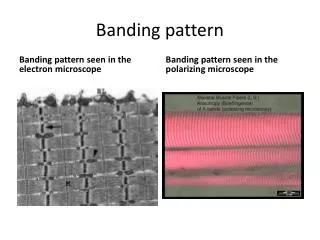
Banding pattern Banding pattern seen in the electron microscope Banding pattern seen in the polarizing microscope
Light bands: I bands (I-LIGHT).Thin filaments only • Dark bands: A bands (A-DARK).overlapping thick and thin filaments. • Z line(Z disc) : dark line in the middle of an I-band.
H-zone : a lighter, central region in the middle of a dark A-band. Only thick filaments. • Sarcomere : runs from Z-line to Z-line (functional contractile unit)
Sliding filament hypothesis of contraction • Nerve impulses carried from brain/spinal cord to muscle by the axon of a motor neuron • Axon meets muscle fiber at the neuromuscular junction(myoneural junction) • Axon releases a chemical neurotransmitter called acetrylcholine, which binds to sarcolemma. • A nerve impulse is generated in sarcolemma and it passes down the t-tubules. As a result the sarcoplasmic reticulum releases Ca++ ions
Sliding filament hypothesis of contraction • Ca++ ions bind to thin filament • This helps thick filament attach to the thin filament • As a result thin filament is drawn further into A band. Myofilaments slides over one another; sarcomere shortens; muscle contracts. • When nerve impulse stops,Ca++ returns to the sarcoplasmic reticulum and the myofilaments slide back to their resting state.