Ischemic Heart Disease
Ischemic Heart Disease. By : Dawit Ayele ( MD,Internist ). Ischemia. •Greek ischein“to restrain” + haima“blood ” •Ischemia occurs when the blood supply to a tissue is inadequate to meet the tissue’s metabolic demands. Causes of Ischemia: Decreased Supply. Hypotension : – Shock….

Ischemic Heart Disease
E N D
Presentation Transcript
Ischemic Heart Disease By :DawitAyele (MD,Internist)
Ischemia •Greek ischein“to restrain” + haima“blood” •Ischemia occurs when the blood supply to a tissue is inadequate to meet the tissue’s metabolic demands
Causes of Ischemia:Decreased Supply • Hypotension: –Shock… •Vascular insufficiency: –Atherosclerosis –Thrombosis –Embolism –Torsion –Compression
Causes of Ischemia: Increased Demand •Increased tissue mass (hypertrophy) •Increased workload (tachycardia, exercise) •Increased tissue “stress” (cardiac dilatation)
Myocardial Ischemia:Occurs when myocardial oxygen demand exceeds myocardial oxygen supply
Effect of Ischemia •Ischemia has 3 principal biochemical components: –Hypoxia (including anoxia) –Insufficiency of metabolic substrates –Accumulation of metabolic waste Effect Depends on: • Severity and Duration of Injury • Cell Type • Microvascular Anatomy
Infarction •Latin infarctus, pp. of infarcire“to stuff” •An infarct is an area of tissue/organ necrosis caused by ischemia •Infarctions often result from sudden reduction of arterial (or occasionally venous) flow by thrombosis or embolism •Infarctions can also result from progressive atherosclerosis, spasms, torsions, or extrinsic
Morphology of Infarcts •Infarcts can be anemic (white) or hemorrhagic (red) •White infarcts occur with arterial occlusion of solid organs •Red infarcts occur with venous occlusion or with arterial occlusions in organs with double or collateral circulation •White infarcts can become hemorrhagic with reperfusion
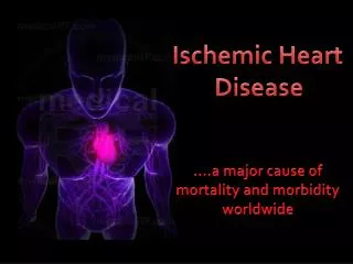
Ischemic Heart Disease (IHD) •Angina pectoris, myocardial infarction, sudden cardiac death, chronic IHD with congestive heart failure •IHD is the leading cause of death in the US and developed countries •Every year in the US, ~1.5 million have an MI and ~600,000 die from ischemic heart disease •Atherosclerosis of the major coronary arteries is responsible for the vast majority of the cases of ischemic heart disease
Acute Myocardial Infarction (MI) •MI indicates the development of an area of myocardial necrosis •MI’s are typically precipitated by an acute plaque change followed by thrombosis at the site of plaque change •Acute plaque changes include fissuring, hemorrhage into the plaque, and overt plaque rupture with distal embolism •Most unstable plaques are eccentric lesions rich in T cells and macrophages, and have a large, soft core of necrotic debris and lipid covered by a thin fibrous cap
Risk Factors • family History • cigarette smoking • diabetes mellitus • hypertension • hyperlipidemia • sedentary life-style • obesity • elevated homocysteine, LP-a ?
Clinical Feature Suggestive features • Different descriptions • Site & radiation: • Typical episode: • Angina equivalents: • Physical exam: risk factors for CAD or cardiac findings of d/t extent
Features suggesting absence of angina pectoris • Pleuritic • Pain localized to tip of one finger • Reproducibility by movement or chest palpation • Constant pain lasting many hours or very brief in seconds • Pain radiating to lower extremities
Differential diagnosis of chest pain according to location where pain starts.
Investigation Supportive • Urine analysis—DM&Renaldis • Lipid profileinine • Hematocrit • Glucose • Creatinine • Thyroid function test • Chest X-ray • Diagnostic • Cardiac enzymes-Cpk,troponin • ECG • Stress test • Coronary angiography
Management plan-should consist of: • 1)Explanation & reassurance • 2)Identification & treatment of aggravating conditions • 3)Adaptation of activity • 4)Treatment of risk factors • 5)Drug therapy for angina and cardiac complications • 6)Consideration of mechanical revascularization Note: IHD needs high index of suspicion , early consultation and referral to cardiac center or hospital with support(Oxygen, NSAIDs, nitroglycerin....)