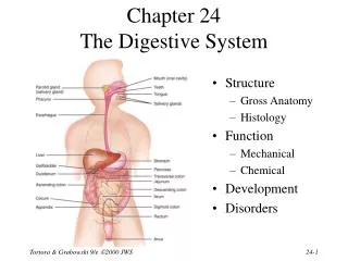
Download
1 / 37
370 likes | 511 Vues
Overview – Cardiac System (Chapter 3 – PW) “ A primer coat”. Sean Collins. Chapter Outline – Level 1. Body Structure and Function Cardiac Evaluation Health Conditions Management. Chapter Outline – Levels 1, 2, 3, 4. Body Structure and Function Cardiac cycle Cardiac Output

E N D
Overview – Cardiac System (Chapter 3 – PW)“A primer coat” Sean Collins
Chapter Outline – Level 1 • Body Structure and Function • Cardiac Evaluation • Health Conditions • Management
Chapter Outline – Levels 1, 2, 3, 4 • Body Structure and Function • Cardiac cycle • Cardiac Output • Factors affecting cardiac output • Preload • Frank-Starling mechanism • Afterload • Cardiac Conduction system • Neural input • Endocrine input • Local input • Cardiac Reflexes • Coronary perfusion • Systemic circulation
Chapter Outline – Levels 1, 2, 3 II. Cardiac Evaluation • Patient history • Physical Exam • Observation • Palpation • Blood pressure • Auscultation
Chapter Outline – Levels 1, 2, 3 II. Cardiac Evaluation • Diagnostic and Laboratory Measures • Oximetry • Electrocardiogram • Complete Blood Cell Count • Coagulation Profiles • Blood Lipids • C-Reactive protein • Biochemical Markers • Natriuretic peptides • ABGs • Chest X Ray • Echocardiograph • Exercise Testing • Cardiac Catherization • Angiography • Electrophysiology studies
Chapter Outline – Level 1, 2 III. Health Conditions • Acute coronary syndrome • Rhythm and conduction disorders • Valvular heart disease • Myocardial and pericardial heart disease • Heart failure
Chapter Outline – Level 1, 2 IV. Management • Revascularization and Reperfusion of the myocardium • Ablation procedure • Cardiac pacemaker implantation and AICD • Life Vest • Valve replacement • Percutaneous aortic valvotomy and transcatheter aortic valve implantation • Cardiac transplantation • Cardiac medications
Chapter Outline – Level 1, 2 IV. Management • Physical Therapy Intervention • Goals • Concepts for the management of patients with cardiac dysfunction
Body structure and function • Cardiac cycle
Chapter Outline – Levels 1, 2, 3, 4 • Body Structure and Function • Cardiac Output • Factors affecting cardiac output • Preload • Frank-Starling mechanism • Afterload • Cardiac Conduction system • Neural input • Endocrine input • Local input • Cardiac Reflexes
Cardiac Evaluation Patient history • Presence of chest pain (see Appendix X for an expanded description of characteristics and etiology of chest pain) • Location and radiation • Character and quality (crushing, burning, numbing, hot), and frequency • Angina equivalents (what the patient feels as angina [e.g., jaw pain, shortness of breath, dizziness, lightheadedness, diaphoresis, burping, nausea, or any combination of these]) • Aggravating and alleviating factors • Precipitating factors • Medical treatment sought and its outcome • Presence of palpitations • Presence of cardiac risk factors (Table 1-4) • Family history of cardiac disease • History of dizziness or syncope • Previous myocardial infarction (MI), cardiac studies, or procedures
Cardiac Evaluation Observation • Facial color, skin color and tone, or the presence of diaphoresis • Obvious signs of edema in the extremities • Respiratory rate • Signs of trauma (e.g., paddle burns or ecchymosis from cardiopulmonary resuscitation) • Presence of jugular venous distention, which results from the backup of fluid into the venous system from right-sided congestive heart failure (CHF) (Figure 1-5) • Make sure the patient is in a semirecumbent position (45 degrees). • Have the patient turn his or her head away from the side being evaluated. • Observe pulsations in the internal jugular neck region. Pulsations are normally seen 3 to 5 cm above the sternum. Pulsations higher than this or absent pulsations indicate jugular venous distention.
Cardiac Evaluation • Palpation • Blood pressure • Auscultation
Cardiac Evaluation Diagnostic and Laboratory Measures • Oximetry • Electrocardiogram • Complete Blood Cell Count • Coagulation Profiles • Blood Lipids • C-Reactive protein • Biochemical Markers
Cardiac Evaluation Diagnostic and Laboratory Measures • Natriuretic peptides • ABGs • Chest X Ray • Echocardiograph • Exercise Testing • Including pharmacological • Cardiac Catherization • Angiography • Electrophysiology studies
Health Conditions • Acute coronary syndrome • Rhythm and conduction disorders • Valvular heart disease • Myocardial and pericardial heart disease All of these roads lead to: Heart failure
Management • Revascularization and Reperfusion of the myocardium • Ablation procedure • Cardiac pacemaker implantation and AICD • Life Vest • Valve replacement • Percutaneous aortic valvotomy and transcatheter aortic valve implantation • Cardiac transplantation • Cardiac medications
Management • Physical Therapy Intervention • Goals • Concepts for the management of patients with cardiac dysfunction