Download

1 / 26
590 likes | 2.06k Vues
Overview of Systemic Lupus Erythematosus. Pediatric Rheumatology Red Team Resident Teaching Series. Systemic Lupus Erythematosus. Episodic, heterogeneous, multisystem autoimmune disease Widespread inflammation of vessels and connective tissues Variable clinical manifestations and course

E N D
Overview of Systemic Lupus Erythematosus Pediatric Rheumatology Red Team Resident Teaching Series
Systemic Lupus Erythematosus • Episodic, heterogeneous, multisystem autoimmune disease • Widespread inflammation of vessels and connective tissues • Variable clinical manifestations and course • characterized by remissions and relapses • Incidence in children: 0.5-0.6/100,000 children/year • 20% of adults have onset in childhood (<19 years old) • Female to male ratio: 9:1 • Uncommon before 4 years of age • More common in African American and Hispanic children • Children have more aggressive disease with more organ involvement than adults • 30% may progress to renal insufficiency depending on treatment • 10 year survival 80-90%
Current Theories Of Pathogenesis In SLE • Etiology unknown • Multiple genes are associated with a predisposition towards developing lupus • Immune dysregulation of B and T cell responses • Immune complex deposition • Abnormalities of complement • Decreased clearance of apoptotic debris • Hormonal imbalance • Environmental triggers including UV B light, infection • Loss of tolerance to chromatin and other autoantigens • Cross reactivity between bacterial and mammalian DNA • Abnormal response to DNA? These factors, acting alone or together, may trigger onset of disease in a genetically predisposed host.
Immune complexdisease • Antibodies can be against self (e.g. nuclear components in SLE) or foreign antigens (i.e. drugs or microorganisms in serum sickness) • Antibodies and antigens combine to form immune complexes • Immune complexes deposit in blood vessels and tissues and activate inflammatory response leading to tissue destruction
ACR 1997 Revised Lupus Criteria Need 4 of 11 criteria for a diagnosis of lupus (sensitivity of 96% and a specificity of 100% in children). If a patient has lupus nephritis, then only a total of 3 of 11 criteria are needed.
2012 SLICC Criteria Click on this link for further details on individual criteria: http://www.rheumtutor.com/2012-slicc-sle-criteria/
Clinical Features of SLE • Constitutional symptoms • Musculoskeletal disease • Mucocutaneousmanifestations • Renal Disease • Central nervous system disease • Cardiopulmonary disease • Hematologic abnormalities • Gastrointestinal involvement
Musculoskeletal Disease • Incidence: 76% • Arthralgias • Arthritis • Non-erosive • Involves small joints of the hands, wrists, elbows, shoulders, knees, ankles • Can be migratory, lasting 24-48 hours • Myalgias/ muscle weakness • Usually proximal
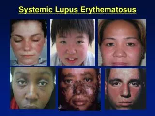
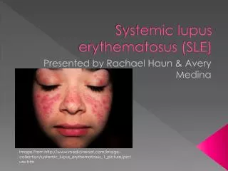
Mucocutaneous Manifestations • Frequency: 76% • Malar rash • Discoid lupus • Vasculitis (purpura, petechiae) • Raynaud’s phenomenon • Nail involvement • Alopecia • Periungual erythema/ Livedoreticularis • Photosensitivity • Oral/ nasal ulcers
Chronic discoid-type malar rash. Discoid lesions tend to be coin-shaped, red and raised with a scaly, sometimes hypopigmented center. This type of lesion can occur in both purely cutaneous (discoid) and systemic lupus. Malar rash: classic butterfly rash, symmetric, sparing nasolabial folds. Can also include the chin and forehead. One third to one half of children have this at presentation. Can be photosensitive.
This is a typical photosensitive eruption involves the face, neck, and "V" of the chest. It occurs after exposure to UV light, and is found in 17-58% of patients with lupus. Crusting has developed following the breakdown of vesicular and bullous lesions. This is livedoreticularis. While not a diagnostic criteria, it is a vasculitic-type rash often seen in individuals with lupus and other vasculitides. Note the mottled, lacey-type pattern.
This is a palatal ulcer. Oral and nasal ulcers occur in 30-40% of lupus patients. Oral ulcers tend to occur on the hard palate, but can occur on the buccal mucosa. They start as hyperemic areas that ulcerate. In the nose, they can precipitate nose bleeds. They are often painless, and are an indicator of active disease. Raynaud’s (single digit): characterized by triphasic color change in an extremity to cold temperature or emotional upset: blanching (white), then cyanosis (blue), then reactive hyperemia (red), accompanied by ischemic pain. To meet a diagnosis of Raynaud’s, the blanching white phase and one other color phase need to be present. Primary Raynaud’s =patients without known autoimmune disease;Secondary Raynaud’s =with autoimmune disease.
Neuropsychiatric Manifestations • Frequency: 20-40% • Difficult to diagnose and treat • Second to nephritis as most common cause of morbidity & mortality • Can occur at any time; even at presentation • Standard lab examinations have not been helpful in diagnosing or managing CNS symptoms • COMMON: Depression, organic brain syndrome, functional psychosis, headaches, seizures, cognitive impairment, dementia, coma • OCCASIONAL:Cerebral vascular accidents (thrombosis or vasculitis), aseptic meningitis, peripheral neuropathy, cranial nerve palsies • RARE:Paralysis, transverse myelopathy, chorea
Cardiovascular Findings • Pericarditis • Most common cardiac manifestation in children • Can be asymptomatic, or represented by chest pain worsened by lying down or deep breathing that is relieved by sitting up and leaning forward. Tamponade is rare but does occur. • Myocarditis • Sterile valvularvegetations (Libman-Sacks endocarditis – see photo below) • Less common in children than in adults. • Subclinical, it is a sterile nodule of fibrinoid necrosis of the collagenous tissue of the valve. • Risk of bacterial endocarditis • Arrhythmias • Corpulmonale • Vasculitis (small vessels) • Atherosclerosis/ Coronary Heart disease • Dyslipoproteinemias
Pulmonary Findings • Incidence: 5-67% • May be subclinical (abnormal PFTs) • Pleuritis • unilateral or bilateral chest pain, exacerbated by deep inspiration • Pleural effusion • Usually small, can be asymptomatic • Pneumonitis • Pulmonary infiltrates, atelectasis, or rales; occurs in 10-15% of children • Pulmonary hemorrhage • Chest pain, hemoptysis, pallor, anemia • Pulmonary hypertension • Restrictive lung disease & diffusion defects most commonly observed abnormalities on PFTs
GI Involvement • Mild LFT elevation--not significant clinically--BUT NEED TO EXCLUDE AUTOIMMUNE HEPATITIS • Colitis • Mesenteric vasculitis • Protein-losing enteropathy • Pancreatitis • Exudative ascites
Leukopenia, especially lymphopenia Neutropenia is rare except in the case of anti-neutrophil antibodies, or in the case of infections Anemia mild to moderate, common, due to chronic disease and mild hemolysis Severe anemia is uncommon (5%), due to immune mediated hemolysis (Coombs +) To meet lupus criteria, the anemia needs to be Coombs positive Thrombocytopenia Due to immune mediated damage mild (100-150K) is common severe (<20K) is uncommon (5-10%) Bone marrow suppression/arrest (aplastic anemia)--very rare, due to antibodies against bone marrow precursors Hematologic Findings
Coagulopathy • Hypocoagulable states: • Anti-platelet antibodies--decreased numbers of platelets or decreased function (increased bleeding time) • Other platelet dysfunction and thrombocytopenia • Anti-clotting factor antibodies • Hypercoagulable states: • Antiphospholipid Antibody Syndrome (APS) • Presence of antiphopholipid antibodies detected by one of the following tests: • Anticardiolipin antibodies • Beta 2-glycoprotein antibodies • Direct Russel Viper Venom Test • Lupus Anticoagulant • Protein C and S deficiencies • Thrombotic thrombocytopenic purpura
Renal Disease • More common and more severe in children than in adults • Most common cause of morbidity & mortality • Can present as proteinuria (including nephrotic range), hematuria, cellular casts, hypertension, decreased GFR <-> renal failure • Glomerulonephritis – occurs in at least 75% • Glomerulonephritis class based on inflammation location and amount, and is predictive of potential for renal damage • Class I+II, III and IV GN (mesangial, focal and diffuse proliferative): result from immune complex deposition from the circulation • Class V GN (membranous): results from in situ formation of immune complexes • Microscopic or gross hematuria • Renal failure (up to 30-50% of children prior to 1980)
Laboratory Findings • Cytopenias (anemia, thrombocytopenia, leukopenia) • Elevated ESR, CRP, Immunoglobulins (particularilyIgG) • Hypoalbuminemia • Proteinuria; RBCs, casts in urine • Decreased creatinine clearance • Low complement levels (C3/ C4) • C4 depression is more specific than C3 depression • Autoantibodies (ANA, APL, Coombs, anti-platelet Ab, rheumotoid factor, etc.) • (Immune complexes)
Antinuclear Antibodies (ANA) • Sensitive but not specific, 95-98% patients positive • Against nuclear components of the cell • Titer specific- up to 10% of population have +ANA w/o disease; also see with infections, medications, malignancy • Subtypes: • dsDNA: high specificity for lupus (over 80%) • ENA (extractable nuclear antigens) • Smith: specific for SLE • U1RNP: associated with MCTD • SSA/SSB: Sjogren’s, neonatal lupus • Histone: drug induced lupus
SLE - Treatment • Treatment is usually based on organ system involvement and the severity of involvement • MILD DISEASE:Rashes, arthritis, leukopenia, anemia, arthritis, fever, fatigue • MODERATE DISEASE:Mild disease + mild organ system involvement such as: mild pericarditis, pneumonitis, hemolytic anemia, mild renal disease • SEVERE DISEASE:Severe, life-threatening organ system involvement • Sunscreen and UV avoidance • Immunizations and infections • Patients on immunosuppressive medications should avoid ALL LIVE VACCINES • Infections should be treated promptly due to immunosuppression and risk of disease flair • Unvaccinated patients exposed to VZV or measles should receive VZIG or IVIG • Physical and occupational therapy
Medications Used in Lupus • NSAIDs • Naproxen – often used with arthritis, mild serositis • Ibuprofen – can cause aseptic meningitis in lupus patients • Antimalarials(usually Hydroxychloroquine) • Used in all patients • Aspirin for patients with APLs • Corticosteroids • Used in all patients except those with very mild disease • Pulse dose • High dose (1-2 mg/kg/day) • Low maintenance dose (5-7.5 mg/day)
Medications Used in Lupus • DMARDs and Immunosuppressives • Azathioprine – hematologic disease, mild NP disease, general steroid-sparing agent • Methotrexate – arthritis, skin disease, general steroid-sparing agent • Cyclosporine/Prograf - nephritis • MMF – nephritis, general steroid-sparing agent • Cytoxan – nephritis, cerebritis, pulm HTN • Biologics • Rituximab • Benlysta • TheurapeuticPlasmaphresis • Stem Cell Transplantation
Emergencies in SLE • Fever: always rule-out infection • Renal disease: uremia, hypertension • Cytopenias: acute hemolytic anemia, thrombocytopenia • CNS: seizures, coma • Pleural effusion (symptomatic)/ pneumonitis/ pulmonary hemorrhage • Pericarditis/ myocarditis/ tamponade • Peritonitis/ pancreatitis/ GI bleed • Raynaud's: digital necrosis • Ocular: retinal vein thrombosis, retinal vasculitis, hemorrhage, edema • Thrombotic events, catastrophic antiphospholipid antibody syndrome (CAPS), thrombotic thrombocytopenic purpura • “Lupus Crisis”
Practice Question • 10-year-old girl with SLE for a health supervision visit. Initially presented at 8 years with ITP, found to have positive ANA, then arthritis and dsDNA. She is currently doing well in school. Malar rash noted on exam. • The MOST useful screening test for other organ involvement is: • A.Coombs test • B.ESR • C.Brain MRI • D.Urinalysis • E.VDRL