Managing Renal and Urologic Infections: Diagnosis, Treatment, and Nursing Care
This resource explores various renal and urologic infections including cystitis, pyelonephritis, and glomerulonephritis. It covers key topics like causes, symptoms, diagnostic methods, and treatment protocols including antibiotics, nursing care, and health promotion strategies. Emphasizing the importance of managing infections related to urinary catheters and pre-existing conditions such as diabetes, this guide serves as an essential tool for healthcare professionals in preventing and treating renal and urologic inflammatory disorders effectively.

Managing Renal and Urologic Infections: Diagnosis, Treatment, and Nursing Care
E N D
Presentation Transcript
Renal & Urologic Problems Nur 302 Unit III
Infection & Inflammatory Disorders • 40% nosocomial, related to cath • Escherichia coli; immunosuppress, DM, mult antibiotics -viral, fungal, parasites • Complicated- coexisting stones, DM, neuro disease, obstructions, catheters • Relapse, reinfections
UTI • Defense mechanisms • Predisposing factors • Sources of UTI- ascending, gram -, nosocomial, abnormal urinary tract
Cystitis • Etiology- anatomic structure & pathologic changes in females, older males, young children • S/S- frequency, urgency, suprapubic pain, foul smelling urine, pyuria, dysuria • Asymptomatic bacteriuria- hematuria, fatigue, anorexia, cognitive changes
Cystitis • Dx: WBC in u/a, urine C&S, gram stain, eval of urinary tract • Meds: Bactrim, Septra, Cipro, Macrodantin, Keflex, Pyridium • Single dose or 1-3 day therapy • UTI with fever, flank pain or chronic- longer therapy • Prophylactic therapy
Nursing Care: Cystitis • Health promotion: identify hi risk pts, teaching fld I, hygiene, empty bladder freq • Prevent nosocomial infection • Increase fld I, avoid bladder irritants, teach drug therapy & s/e, teach s/s UTI • Follow up care with urine C&S, can relapse in 1-2 weeks
Acute Pyelonephritis • Acute or chronic inflamm of renal pelvis or parenchyma of kidney • Infection ascends from lower urin tract • Often, preexisting factor • Chronic pyelonephritis- starts in medulla, spreads to cortex, heals, fibrosis, scars
Acute Pyelonephritis • S/S: mild lassitude, s/s cystitis, sudden fever, chills, vomiting, malaise, flank pain, costovertebral tenderness on affected side • CBC- leukocytosis, incr banded neutrophils, u/a- pyuria, bacteriuria, hematuria, wbc casts • Bacteremia, septic shock
Pyelonephritis • Dx- u/a, C&S, Gram’s stain, WBC, blood C&S, flank pain, ultrasound, CT scan • Consider contributing factors, IVP later • Antibiotics 14-21 days, rx of relapse with 6 wks or prophylactic antibiotics • Evaluate with urine C&S
Nursing Care: Pyelonephritis • Health Promotion: stress reg med care • Teach: continue med, importance of follow-up urine C&S, s/s relapse, drink 8 glasses water minimum, rest • Treat s/s- hyperthermia, pain, see NCP 46-1
Chronic Pyelonephritis • Predisposing factors: chronic UTIs, obstruction, neurogenic bladder, vesicouretal reflux • Chronic inflammation & scarring, renal pelvis & calyces dilated, deformed • Destruction of nephrons->renal insuff • End stage chronic renal failure
Urethitis • S/S same as cystitis, discharge, urethra tender, bacteria in edematous urethral tissue & don’t appear in u/a • Causes: viral, Trichomonas & monilial infection, Chlamydia & gonorrhea • Split urine C&S, C&S discharge • Rx: antibiotics, sitz bath, proper cleansing, no vaginal deodorant, avoid sex
Urethral Syndrome • Acute urethral syndrome: dysuria, urgency, frequency with bacteriuria • Bacteriuria: E. coli, enterococci, staph • Chlamydia, gonorrhea if few bacteria • R/O vaginitis • TX depends on cause
Renal Tuberculosis • Secondary to TB of lung, onset 5-8 later • Initially, no s/s, low fever, fatigue • Lesions ulcerate, spread to bladder-> s/s cystitis; may calcify-> lumbar & iliac pain, hematuria, renal colic • Dx: urine C&S, IVP • Complications: strictures, scarring renal parenchyma, renal failure
Glomerulonephritis • Inflammation of glomerulus with tubular, interstitial & vascular changes • Immunologic, antibody induced injury • Anti-GBM antibodies stimulated by structural alteration of GBM or reaction to virus & results in deposits in GBM • Antibodies react with nonglomerular antigens & randomly deposited, look “lumpy bumpy”
Glomerulonephritis • Accumulation of antibody, antigen, compliment in glomeruli-> tissue injury • Compliment activation-> leukocytes, release of histamine & vasoactive amines, clotting mechanism activated • S/S: hematuria, u/a has WBC, RBC, casts, proteinuria, elev BUN, creatinine
Acute Poststreptococcal Glomerulonephritis (APSGN) • 5-21 days after skin or throat infection • Group A Beta hemolytic streptococci • Antibodies to strep develop->inflam-> decreased filtration of metabolic waste, & increased permeability protein • S/S: none or generalized edema, oliguria, hi BP, “rusty” hematuria, proteinuria, flank pain
APSGN • Dx: H&P, u/a, CBC, BUN, creat, albumin, ASO titer, renal biopsy • Nsg Care: rest, Na & fld restriction, diuretics, antihypertensive meds, lo P diet, antibiotics if have strep • Encourage early tx of sore throat & skin lesions, teach good hygiene & take all antibiotics
Rapidly Progressing Glomerulonephritis (RPGN) • Renal failure occurs within weeks • Occurs as compliment of inflammatory disease, complication of systemic disease (Lupus), idiopathic, or assoc with drugs (PCN) • Manage fld overload, hi BP, uremia • Dialysis & transplant but RPGN can reoccur
Nephrotic Syndrome • Causes: glomerulonephritis, infections, multisystem diseases, neoplasms, allergens • S/S: periph edema, proteinuria, hi lipids, lo albumin, ascites, anasarca, altered immune response -> infection, hypocalcemia, loss of clotting factors-> hypercoagulability, thrombus formation esp R renal vein, PE
Nephrotic Syndrome • Tx: relieve edema, control disease • ACE inhibitors, NSAIDs, lo Na diet, loop diuretic • Lipid lowering agents • Anticoagulants if thrombus • Corticosteroids & Cytoxin
Nursing Care • Assess edema: daily wt, I&O, measure girth • Skin care, prevents trauma->weeping • Monitor diuretic therapy, labs • Lo protein-> malnourished, anorexic, lo Na & P diet; assess dietary needs, sm freq feedings • Prevent infection • Altered body image- psychol support
Obstructive Uropathies • Causes- intrinsic, extrinsic, functional • System above level of obstruction is affected • Location, duration, pressure, urinary stasis, infection affect severity of effects • Obstruction distal to prostate or bladder neck->mucosal scarring & slower stream • Obstruction at prostate or bladder neck-> tabeculation, diverticuli, incr pres, reflux
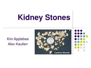
Urinary Tract Calculi • Stone formation: genetic, metabolic, dietary, climatic, lifestyle, occupational • Calculus- stone & lithiasis- formation • Types of stones- see table 46-12 • S/S occur where stone causes obstruction to urine flow; severe abd or flank pain, hematuria, renal colic, n/v, UTI s/s • Passing stone- intense, colicky pain, mild shock with cool, moist skin
Urinary Tract Calculi • Dx: history, u/a, C&S, IVP, retrograde pyelogram, ultrasound, cystocopy, abd x-ray, CT, urine & serum levels of stone metabolites, BUN, Creat, urine ph • Manage acute attack- treat pain, infection, obstruction • Eval of composition of stone & prevent further formation of stones
Urinary Tract Calculi • Indications for endourologic, lithotripsy or surgery • Cystoscopy • Cystolitholapaxy • Cystoscopic lithotripsy • Ultrasonic, laser or electrohydraulic lithotripsy • Percutaneous nephrolithotomy
Nursing Care • Prevention- esp pts on BR with urinary stasis, incr fld I minimum 2L/day, diet restrictions purine, oxalate calcium • See NCP 46-2 • Strain all urine • Pain management • Teaching- diet, flds, meds, test urine ph
Strictures • Congenital or acquired • Occur at bladder neck, urethra, ureters • Causes: trauma, gonorrhea, urethral instruments, chronic infections, radiation, retroperitoneal abscess • Treatment : dilitation with catheter, drainage with catheter, surgery
Renal Trauma • Blunt trauma common- car accidents, sports, falls with injury to flank, abdomen or back • Penetrating – gunshots, stabbing • Dx: history, hematuria, u/a, IVP with cystogram, ultrasound, CT, MRI • Nsg Care: Monitor I&O, hematuria & nephrotoxic antibiotics, pain, s/s shock
Nephrosclerosis • Sclerosis of small arteries & arterioles-> decr bld flow-> patches of necrosis-> destruction of glomeruli & fibrosis • Benign nephrosclerosis due to hi BP, & arteriosclerosis • Accelerated or malignant due to malig hi BP, diastolic >130-> renal insuffic-> renal failure eventually • Prevention & rx: treat hypertension
Renal Artery Stenosis • Partial occlusion renal a. due to atherosclerosis or fibromuscular hyperplasia • Dx: renal arteriogram • Rx: control BP, angioplasty, stints, surgical anastomoses bet kidney & spleenic artery or aorta
Polycystic Renal Disease • Genetic, latent, s/s appear age 30-40 • Cortex & medulla filled with cysts • S/S when cysts enlarge- abd or flank pain, palpable enlarged kidneys, UTI, hi BP, hematuria, 50% develop renal fail. • Dx: H&P, CT, IVP, ultrasound • Rx: prevent UTI, nephrectomy, genetic counseling
Medullary Cystic Disease • Hereditary • Recessive form-> renal fail. before 20 • Dominant form-> renal failure after 20 • Affects ability to concentrate urine • Polyuria, severe anemia, renal failure, metabolic acidosis, poor Na concentration
Renal Problems in Metabolic & Connective Tissue Diseases • Diabetic neuropathy • Gout • Amyloidosis • Systemic Lupus Erythematosus • Scleroderma
Renal Tumors • Arise from cortex or pelvis, benign or malignant- adenocarcinoma • Risk factors- smoking, exposure to asbestos, gasoline, cadmium, phenacetin containing analgesics • S/S: wt loss, anemia, weakness, gross hematuria, flank pain, palpable mass • Metastasis- lungs, liver, long bones, renal vein & vena cava
Renal Tumors • Dx: IVP with nephrotomography, CT, MRI, angiogram, needle aspiration • Staging- Robson’s system • Tx: nephrectomy, radiation palliatively, no chemo available, biologic therapy
Bladder Cancer • Most common- transitional cell carcinoma, papillomatous • Risk factors: smoking, dyes used in rubber & cable industry, phenacetin-containing analgesics, women tx with Cytoxin for cervical cancer • Chronic stones->risk for squamous cell bladder cancer
Bladder Cancer • S/S: gross & painless hematuria, also dysuria, freq, urgency • Dx: urine for cytology, bladder tumor antigens, IVP, ultrasound, MRI • Definite dx by cystoscopy & biopsy • Jewett-Strong-Marshall classification: superficial, invasive, metastatic
Surgery: Bladder Cancer • Transurethral resection with fulgaration • Laser photocoagulation • Open loop resection with fulgaration • Post-op care: increase fld I, I&O, avoid alcohol, analgesics, sitz baths, psychol support, reg follow ups & cystoscopies • Radical cystectomy
Tx Bladder Cancer • Radiation therapy • Chemotherapy: Vinblastine, Platinol, Adriamycin, Methotrexate • Intravesicular therapy: instill chemo into bladder via catheter • S/E: irritating voiding, hemorrhagic cystitis, decr WBC & platelets
Urinary Incontinence • Stress incontinence • Urge incontinence • Overflow incontinence • Reflux incontinence • Incontinence after trauma or surgery • Functional incontinence
Neurogenic Bladder • Bladder dysfunction from CNS neurologic disorder • Tumors, spinal cord injury, CVA, MS, diabetic neuropathy • Failure to store, empty or both • Dysfunction of bladder or urethra • Location- whether it affects brain or spinal cord
Causes of Urinary Retention • Antihypertensives- Aldomet, Apresoline • Antiparkinsonian- Levodopa • Antihistamines • Anticolinergics- Atropine • Antispasmodics • Sedatives & spinal anesthesia • Urethral obstruction • Psychological