Download
1 / 136
1.37k likes | 1.67k Vues
Recombination, Bacteriophages, and Horizontal Gene Transfer. 2005. Bacterial Conjugation. transfer of DNA by direct cell to cell contact discovered 1946 by Lederberg and Tatum. F + x F – Mating. F + = donor contains F factor F – = recipient does not contain F factor

E N D
Recombination, Bacteriophages, and Horizontal Gene Transfer 2005
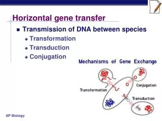
Bacterial Conjugation • transfer of DNA by direct cell to cell contact • discovered 1946 by Lederberg and Tatum
F+x F– Mating • F+ = donor • contains F factor • F– = recipient • does not contain F factor • F factor replicated by rolling-circle mechanism and duplicate is transferred • recipients usually become F+ • donor remains F+
F factor • The F factor can exist in three different states: • F+ refers to a factor in an autonomous, extrachromosomal state containing only the genetic information described above. • The "Hfr" (which refers to "high frequency recombination") state describes the situation when the factor has integrated itself into the chromosome presumably due to its various insertion sequences. • The F' or (F prime) state refers to the factor when it exists as an extrachromosomal element, but with the additional requirement that it contain some section of chromosomal DNA covalently attached to it. A strain containing no F factor is said to be "F-".
Gene transfer and recombination • Genes are transferred in a linear manner • The F factor integrates into chromosomes at different points and its position determines the O site
Hfr Conjugation • Hfr strain • donor having F factor integrated into its chromosome • both plasmid genes and chromosomal genes are transferred
Hfr • Special class of F+ strains • This was discovered because this strain underwent recombination 1000x more frequently than F+ strains • In certain Hfr strains certain stains are more likely to recombine than others. • The nonrandom pattern of gene transfer was shown to vary from Hfr strain to Hfr strain
Interrupted mating • Wollman explained the cells that are different between F+ and Hfr. To facilitate the recovery, the Hfr was sensitive to antibiotics and the F+ wasn’t. • The cells were separated at intervals of 5 minutes is the F factor
Hfr x F– mating Figure 13.14b
Recombination • Exogenote • Exogenote
Mating • The two strains were mixed • There were incubated. • At intervals of 5 minutes, samples were taken of the F- cells • The cells were centrifuged so that they would know which genes were transferred. • The distance between genes was measured by the time that it took for the genes to be transferred. • During the first five minutes, the strains were mixed there was no recombination
F+x F– mating • In its extrachromosomal state the factor has a molecular weight of approximately 62 kb and encodes at least 20 tra genes. It also contains three copies of IS3, one copy of IS2, and one copy of a À sequence as well as genes for incompatibility and replication.
F’ • In 1959 during his experiments with the Hfr strains of E. coli Adelberg discovered that the F factor could lose its integrated status and revert to its F+ status. • When this occurred, the F factor carries along several adjacent bacterial genes. • When you have the F factor + bacterial genes – the condition is known as the F’
F Conjugation integrated F factor • F plasmid • formed by incorrect excision from chromosome • contains 1 genes from chromosome • F cell can transfer F plasmid to recipient chromosomal gene Figure 13.15a
Merozygotes • When the F’ is then transferred to another bacterium • The bacterium may contain genomic copies of a gene as well as an additional copy of the gene in the F’. • As a result the situation is a partial diploid • Merozygotes have been extremely beneficial in the study of gene regulation
Interrupted mating Figure 13.22a
Hfr mapping • used to map relative location of bacterial genes • based on observation that chromosome transfer occurs at constant rate • interrupted mating experiment • Hfr x F- mating interrupted at various intervals • order and timing of gene transfer determined
Recombinants • Map distance can be determined by replating the resulting colonies on agar • For example leu+ exconjugants by plating them on medium containing no leucine but containing methionine and arginine
Map distance • The map distance is equal to the % recombination
Tra Y • Characterization of the Escherichia coli F factor traY gene product and its binding sites • WC Nelson, BS Morton, EE Lahue and SW Matson Department of Biology, University of North Carolina, Chapel Hill 27599.
Tra Genes • Tra Y gene codes for the protein binds to the Ori T • Initiates the transfer of plasmid across the bridge between the two cells • Tra I Gene is a helicase responsible for the conjugation • strand-specific transesterification (relaxase)
Conjugative Proteins • Key players are the proteins that initiate the physical transfer of ssDNA, the conjugative initiator proteins • They nick the DNA and open it to begin the transfer • Working in conjunction with the helicases they facilitate the transfer of ss RNA to the F- cell
DNA Transformation • Uptake of naked DNA molecule from the environment and incorporation into recipient in a heritable form • Competent cell • capable of taking up DNA • May be important route of genetic exchange in nature
Streptococcus pneumoniae nuclease – nicks and degrades one strand DNA binding protein competence-specific protein
Artificial transformation • Transformation done in laboratory with species that are not normally competent (E. coli) • Variety of techniques used to make cells temporarily competent • calcium chloride treatment • makes cells more permeable to DNA
Transformation mapping • used to establish gene linkage • expressed as frequency of cotransformation • if two genes close together, greater likelihood will be transferred on single DNA fragment
Bacteriophages Microbial Genetics
Diversification of Escherichia coli genomes: are bacteriophages the major contributors? Makoto Ohnishi – Trends in Microbiology • E. coli is a diverse species • 4.5 – 5.5 MB • E. coli strains are commensals of higher vertebrates, but some are pathogenic • There are 5subtypes of the diarrheagneic strainsd • The pathogenicity of the strains has been traced to a subtype that retains a large segment of virulence factors or pathogenicity islands
E. Coli O 157 • Sixteen sections of this pathogenic strain differ from the lab strain • These are subtype specific • Within sections of the DNA and these large segments • The G-C content varies from the lab strain • The 4.1 kb common backbone sequence mainly represent the DNA that RE. coli possesses from a common ancestor.
E. coli • There are 98 copies of IS elements within this section as well as genes enxoding hemolysins, proteases, and other virulence factors. • More interesting O 157 also contains 18 remnants of prophages
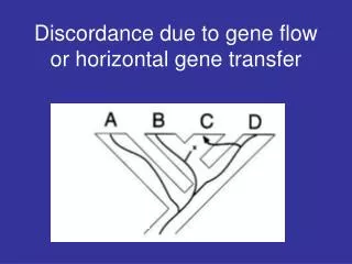
Horizontal gene transfer • Clearly this plays a central role in the diversity of E. coli • Among the 18 prophage remnants on O157 – 12 resemble lambda pahge • They all contain a variety of deletions and or insertions • Some of the phages are so similar that they contain a 20 kb segment tat is identical.
Recombinant phages • It is believed that the phages have undergone recombination and diversification • Recombination could occur with in a single cell • It could occur as the result of recombination
Virulence and Strptococcus pyogenes • Streptococcal pyrogenic exotoxins(SPE) contribute to the diverse symptoms of a streptococcal infection. • These antigens compare to Staphylococcal antigens of the same type. • The A + C genes coding for these toxins were horizontally transferred from strain to strain by a lysogenic bacteriophage. • In addition the genes contributed by the phages produce hyaluronidase, mitogenic factor, and leukocyte( WBC) toxins
Streptococcus pyogenes • There are 15 prophages that have been identified in E. coli • These prophages belong to the group Siphoridae • All but one of these produce a toxin • In both strep and staph – the prophage is found at the site of recombination
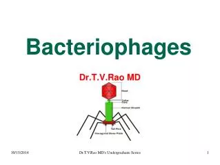
Bacteriophages • Bacterial viruses • Obligate intracellular parasites • Inject themselves into a host bacterial cell • Take over the host machinery and utilize it for protein synthesis and replication
T- 4 Bacteriophage • Ds DNA virus • 168, 800 base pairs • Phage life cycles studied by Luria and Delbruck
Bacteriophage structure(con) • Most bacteriophages have tails • The size of the tail varies. • It is a tube through which the nucleic acid is injected as a result of attachment of the bacteriophage to the host bacterium • In the more complex phages the tail is surrounded by a contractile sheath for injection of the nucleic acids
Bacteriophage structure • Many bacteriophages have a base plate and tail fibers • Some have icosahedral capsids • M13 has a helical capsid
Bacteriophage structure(con) • Most bacteriophages have tails • The size of the tail varies. • It is a tube through which the nucleic acid is injected as a result of attachment of the bacteriophage to the host bacterium • In the more complex phages the tail is surrounded by a contractile sheath for injection of the nucleic acids