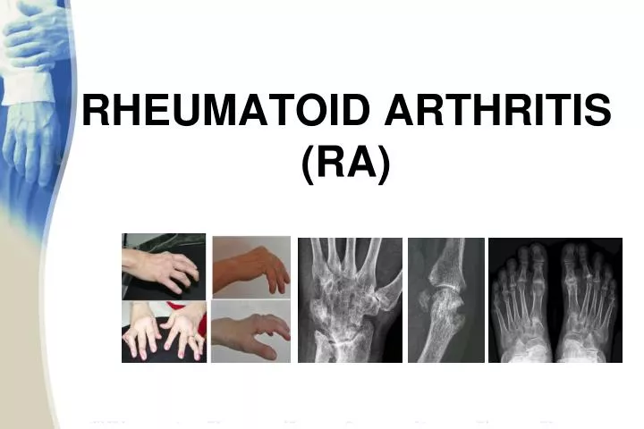
RHEUMATOID ARTHRITIS (RA)
1.22k likes | 2.64k Vues
RHEUMATOID ARTHRITIS (RA). Overview. Definition Immuno -pathogenesis Clinical findings (articular and systemic manifestations) Investigations Assessment & monitoring Management. RHEUMATOID ARTHRITIS. Chronic multisystemic inflammatory disease of unknown etiology

RHEUMATOID ARTHRITIS (RA)
E N D
Presentation Transcript
Overview • Definition • Immuno-pathogenesis • Clinical findings (articular and systemic manifestations) • Investigations • Assessment & monitoring • Management
RHEUMATOID ARTHRITIS • Chronic multisystemicinflammatory disease of unknown etiology • Affects the synovial membranes of multiple joints (diarthrodial joints) • Female : Male ratio 3:1 • Most frequent during 4th and 5th decade • Affects approximately 1% of the adult population worldwide
DEFINITION chronic immune inflammatory disorder, with still unknown aetiology, characterized by: • Articular manifestations: chronic destructive and deforming arthritis affecting small joints in a bilateral and symmetrical pattern; • Systemic manifestations: cardio-vascular, respiratory, renal, neurologic, ocular; • Progressive irreversible articular damage; • Significant functional disability and impaired quality of life
IMMUNO-PATHOGENESIS Proliferative synovitis(pannus) Inflammation, (neo)angiogenesis, tissue damage
NORMAL SYNOVIAL JOINT Type A cell: mphage like, protective role Synovial Membrane: only 1-2 cells thick Type B cell: fibroblast like, produce matrix and synovial fluid Vessel Synovium Membrane Articular cartilage Lamellar bone Subchondral bone
Chondrocytes themselves Proliferating synovial lining => Pannus IL-1 and TNF-alpha also stimulate production of metalloproteinase by Chondrocytes of the articular cartilage. In response to these cytokines, chondrocytes decrease type II collagen and proteoglycan synthesis and increase synthesis of metalloproteinases that contribute to the degradation of collagen and proteoglycans. Proliferating synovial lining comes in contact with the cartilage matrix and bone there is degradation of the cartilage and erosion of the bone surface (by matrix metalloproteinases and other proteases produced by synovial cells) cartilage bone Neutrophils in Synovial Fluid Osteoclasts The main inflammatory cells of the synovial fluid are neutrophils. Cytokines such as transforming growth factor beta (TGF-beta) and interleukin 8 (IL-8) attract neutrophils. Neutrophils may undergo degranulation and cause some damage to surrounding tissues. Osteoclasts may be activated by inflammatory mediators including IL-1, TNF and PGE2 DESTRUCTION OF JOINT CARTILAGE AND BONE
CENTRAL ROLE OF TNFα IN RA Kirwan JR. J Rheumatol. 1999;26:720-725.
EARLY CHANGES IN RA Thickened Synovium Lymphocytic Infiltrate Neovascularization Major type of cells in synovium are T-cells and macrophages whereas in synovial fluid are neutrophils
RA IS CHARACTERISED BY SYNOVITIS AND JOINT DESTRUCTION NORMAL RA Inflamed synovial membrane Synovial membrane • Major cell types: • T lymphocytes • macrophages Pannus Cartilage • Minor cell types: • fibroblasts • plasma cells • endothelium • dendritic cells Synovial fluid • Major cell type: • neutrophils Capsule Cartilage thinning Adapted from Feldmann M, et al. Annu Rev Immunol. 1996;14:397-440.
CLINICAL MANIFESTATIONS • Articular findings: rheumatoid hand and foot; C1-C2 subluxation • Extra-articular findings: cardio-vascular (rhythm troubles, early accelerated atherosclerosis, vasculitis), respiratory (diffuse interstitial fibrosis, nodules, pleurisy), ocular (sicca syndrome), neurologic (peripheral & entrapment neuropathy), renal (amyloidosis), rheumatoid nodules, myositis, osteoporosis • Chronic evolution with flares ACR 1987 Diagnostic Criteria ACR and EULAR 2010 Classification Criteria ACR, American College of Rheumatology EULAR, European League Against Rheumatism
RHEUMATOID ARTHRITIS • Clinical presentation • usually presents insidiously; • prodromal syndrome of malaise, weight loss and vague periarticular pain and stiffness may be seen • less commonly, the onset is acute, triggered by a stressful situation such as infection, trauma, emotional strain or in the postpartum period. • the joint involvement is characteristically symmetric with associated stiffness, warmth tenderness and pain
RHEUMATOID ARTHRITIS • Clinical Features • the stiffness is characteristically worse in the morning and improves during the day; its duration is a useful indicator of the activity of the disease. • the usual joints affected by rheumatoid arthritis are the metacarpophalangeal joints, the PIP joints, the wrists, knees, ankles and toes. • Entrapment syndromes may occur especially carpal tunnel syndrome
RHEUMATOID HAND • Swelling of the RC, MCF, PIP joints • Fusiform swelling fingers: synovitis of PIP joints, causing them to appear spindle-shaped. DEVIERE CUBITALA DEGETE POLICE “IN Z”
RHEUMATOID ARTHRITIS • After months to years, deformities can occur; the most common are • ulnar deviation of the fingers • swan neck deformity, which is hyperextension of the distal interphalangeal joint and flexion of the proximal interphalangeal joint • boutonniere deformity, which is flexion of the distal interphalangeal joint and extension of the proximal interphalangeal joint • valgus deformity of the knee
RHEUMATOID HAND • Fingers in “swan-neck” • Fingers in “ boutonniere” • artritismutilans Swan-neck deformity” DEGETE “IN GAT DE LEBADA” Boutonniere deformity Rheumatoid nodules Artritismutilans
RHEUMATOID ARTHRITIS Saurabh Garg
RHEUMATOID FOOT Hammer toes Complex deformity of the foot Subluxation of the metatarsal heads Triangular foot
EXTRA-ARTICULAR MANIFESTATIONS • General: fever, lymphadenopaty,weit loss, fatigue • Dermatologic: rheumatoid nodules, vasculitis • Ocular manifestations: Keratoconjunctivitis sicca, episcleritis, scleritis, choroid and retinal nodules • Pulmonary manifestations: pleural involvement, fibrosing alveolitis , obliterative bronchiolitis • Felty’s Syndrome: RA with splenomegaly and neutropenia • Cardiac involvement: Constrictive pericarditis, myocarditis, coronary vasculitis, nodules on valves • Renal involvement: secondary amyloidosis • Neurologic manifestations: Mononeuritismultiplex, entrapment neuropathies, peripheral neuropathies • Hematologic manifestations: anemia, thrombocytosis
RHEUMATOID NODULES Subcutaneus nodules Occur 20-35% of RA patients in severe, active disease with RF positive localisation: on the extensor surface of the forearms, in the olecranon bursa, over joints, and over pressure points, like sacrum and occiput characteristic histology: central area of fibrinoid necrosis surrounded by a zone of palisades of elongated histiocytes and a peripheral layer of cellular connective tissue
VASCULITIS In RA patients with long standing disease, significant joint involvement, high titers RF, and nodules Clinical aspects: palpable purpura,small infarcts of digital pulp,visceral arteritis
OCULAR MANIFESTATIONS keratoconjunctivitissicca (30%) iritis/iridociclitis episcleritis/scleritis scleromalaciaperforans retinianvasculitis EPISCLERITIS SCLERITIS SCLEROMALACIA PERFORANS
RHEUMATOID ARTHRITIS • Labs: inflammatory syndrome: • The ESR is elevated both in the acute and chronic phases of the disease • C reactive protein • a moderate anemia is often present which is usually hypochromic normocytic • the white count is normal or slightly increased but leukopenia may occur, often in presence of splenomegaly (e.g., Felty’s syndrome) • the platelet count is often elevated in proportion to the degree of joint inflammation
RHEUMATOID ARTHRITIS • Labs: immune syndrome: • Rheumatoid factor is an autoantibody directed against the constant region (Fc) of IgG. High titers of rheumatoid factor are associated with severe disease. Rheumatoid factor is also found in other diseases like syphilis, sarcoidosis, infective endocarditis, TB, leprosy, parasitic,SLE, hepatitis, infections; in advanced age and in asymptomatic relatives of patients with rheumatoid disease. • Antinuclear antibody are seen in 20% of patients with rheumatoid arthritis, though their titer is lower than in SLE • Anticycliccitrullinated peptide antibody are the most specific for RA; they are correlate strongly with erosive disease.
RHEUMATOID ARTHRITIS • Labs • joint fluid examination is valuable. The fluid is translucent to opaque and has between 3000 and 50,000 WBCs /microL. There are 50% or more polymorphonuclear leukocytes. The culture is negative. • X-ray • of all the laboratory tests, x-ray changes are most specific for rheumatoid arthritis. However, they are not sensitive and usually are negative during the first 6 months of the disease
RHEUMATOID ARTHRITIS • X-rays • The earliest changes occur in the wrist or feet and consist of soft tissue swelling and juxta-articular demineralization. • Later, diagnostic changes consisting of joint space narrowing and erosions develop. The erosions are first seen at the ulnar styloidand at the juxta-articular margin, where the bony surface is not protected by cartilage. • Diagnostic changes also occur in the cervical spine with C1-2 subluxation, but this can take several years to develop.
geodes erosion
C1-C2 Subluxation
RHEUMATOID ARTHRITIS • 1987 American College of Rheumatology Revised criteria for the diagnosis of Rheumatoid Arthritis: • At least four of the following • 1. Morning stiffness > 1hour • 2. Synovitisin three joints simultaneously • 3. Synovitisin wrist or hand, MCP or PIP joints • 4. Symmetrical arthritis (some joint areas on both sides of the body) • 5. Rheumatoid nodules • 6. Serum rheumatoid factor • 7. Radiographic changes typical of Rheumatoid Arthritis (erosions are the patognomonicalsignes) • To be classified as having RA a patient must meet 4 or more criteria
PROBLEMS WITH OLD ACR CRITERIA • Work best in longstanding RA • But DMARDs work best in early RA and the goal is to prevent development of damage • Need criteria addressing earlier diagnosis given the benefits of early treatment • Need to include ACPA (CCP) • Balance with need for use in low resource settings where CCP not available • Goal of new criteria: predict who should be treated with DMARDs
NEW ACR/EULAR PROPOSED CRITERIA • Initial screen: • 1+ swollen joints (if no, not RA) • Better explained by other dz? (if yes, not RA) • Typical RA erosion on X-ray? (if yes, RA) • Next step: • Pattern of joint involvement (more points for more joints and small joints) • Serology (RF and/or CCP, negative, low, high) • Duration (<6 wk, 6+ wk) • ESR and/or CRP (both normal vs. one abnormal)
2010 ACR/EULARClassification Criteria for RA ≥6 = definite RA What if the score is <6? Patient might fulfill the criteria… Prospectively over time (cumulatively) Retrospectively if data on all four domains have been adequately recorded in the past
ASSESSMENT AND MONITORING • RA activity: Disease Activity Score, DAS28 • Disability and quality of life: Health Assessment Questionnaire, HAQ • Response to treatment: EULAR, ACR criteria • Remission • Negative prognostic factors
ACR GUIDELINES FOR MANAGEMENT • Summarize evidence for DMARDs and biologics in different settings • Incorporate the following in treatment decisions • Disease duration (<6mo, 6-24, >24 mo) • Disease activity (low, moderate, high) • Features of poor prognosis Saag KG et al. Arthritis Rheum 2008;59:762
MEASUREMENT OF DISEASE ACTIVITY: DAS28 AS EXAMPLE DAS28 = 0.56 * sqrt(tender28) + 0.28 * sqrt(swollen28) + 0.70 * ln(ESR) + 0.014 * GH • Includes: • Tender joint count • Swollen joint count • ESR (or CRP in different version) • GH: Patient global disease activity assessment • Categorized: as low (<3.2), moderate (3.2-5.1), or high (>5.1)
DAS 28 28 joints counted