Download

1 / 41
520 likes | 4.4k Vues
Echocardiography in the Diagnosis of Mitral Valve Prolapse 2-D Criteria At one time the diagnosis of mitral valve prolapse was based on M-mode criteria.

E N D
2-D Criteria • At one time the diagnosis of mitral valve prolapse was based on M-mode criteria. • With the application of 2-D echocardigraphy, which visualizes mitral valve motion in real time, however, this method has a higher sensitivity in the diagnosis of mitral valve prolapse
2-D Criteria • The mitral valve is shaped like a saddle in 3 dimensions, with its high points, farthest from the apex, located anteriorly and posteriorly in the long-axis view.
2-D Criteria • A 4 chamber view through such a structure can normally show leaflets apparently budging upward relative to the low points of the annular saddle without any leaflet disease or distortion.
2-D Criteria • This particularly applies to superior bowing of the anterior mitral leaflet which heads superiorly towards the aortic root in the anterior posterior view but more apically towards the central fibrous body of the heart in the apical 4 chamber view.
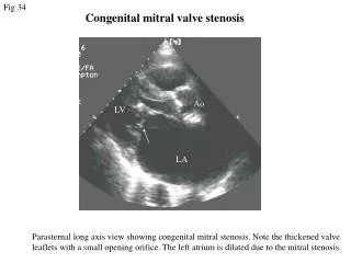
Therefore, the diagnosis of mitral valve prolapse is based on the parasternal long-axis view Anterior or posterior mitral leaflet prolapse is considered present when any portion of the leaflet protrudes beyond the mitral annular plan >2mm 2-D Criteria
2-D Criteria • The diagnosis is more certain when the mitral leaflets are thickened (myxomatous). • When leaflet thickness is >5mm measured in the parasternal long axis view during diastole or diastolic leaflet thickness is > 1.4 times the thickness of the posterior wall of the aorta
2-D Criteria • The size of the LA should be measured at end-systole. • This is an inner edge to inner edge measurement (normal 2.3 to 2.8cm)
2-D Criteria • Other associated conditions should be ruled out such as Marfan syndrome
M-mode Criteria • Because of the excellent temporal resolution of M-mode, it can still be used in conjunction to the 2-D diagnosis of MVP • The leaflets will appear thick, redundant. The thickness of the mitral valve leaflets can be determined by measuring the thickness of the anterior and/or posterior leaflets in mid-diastole. • > 5mm which correlates with 2-D is considered to be evidence of myxomatous leaflets
M-mode Criteria • Mid to late systolic “sagging” back of the anterior, posterior, or both leaflets > 2mm from the C-D points
M-mode Criteria • Holosystolic (pansystolic) “sagging” back of the anterior, posterior, or both mitral valve leaflets > 3mm from the C-D points
M-mode Criteria • LA dilatation (significant mitral regurgitation)
Doppler/color Doppler Criteria • In color-flow imaging of mitral regurgitation, the area of the regurgitant jet relative to the size of the LA is most predictive of regurgitant severity determined with angiography. • Color-flow imaging of valvular regurgitation depends on the gain setting, pulse repetition frequency, field depth, direction of jet, and loading conditions.
Doppler/color Doppler Criteria • A flow jet directed against the atrial wall appears smaller than a free jet of the same regurgitant volume (Coanda effect). Therefore, jet size on color-flow imaging should be interpreted in the context of jet geometry and the surrounding solid boundaries
Doppler/color Doppler Criteria • On Doppler, the velocity of MR tends to be lower <5m/sec with increasing severity because the increase in LA pressure reduces the transmitral systolic gradient, unless the LV pressure is markedly increased.
Doppler/color Doppler Criteria • In severe MR, there may be systolic flow reversal in the pulmonary veins
Doppler/color Doppler Criteria • Most recently, vena contracta width by color-flow mapping correlates well with other quantitative measures for mitral regurgitation severity. • Vena contracta is the narrowest portion of the MR jet downstream from the orifice. • A biplane vena contracta width >0.5cm. This would be considered hemodynamically significant MR.
Doppler/color Doppler Criteria • In the case of prolapse, MR is often late systolic • Depending on the leaflet or leaflets that are affected, will determine the direction of the mitral regurgitant jet • If there was an posterior MVP which direction would the MR jet go? • If there was a anterior MVP which direction would the MR jet go? • If both leaflets prolapsed, which direction would the MR jet go?
Doppler/color Doppler • E.g., posterior prolapse, anterior direction, anterior prolapse, posterior direction, both leaflets, central direction
Echocardiography in the Diagnosis of Flailed Mitral Valve • A flail mital valve is best detected with 2-D echocardiography • Extension of part of the valve into the left atrium in SYSTOLE can be readily noted. • What views would show a flailed leaflet the best?
2-D • It is important to try and identify the reason for the flailed MV leaflet • Ruptured chordae • Ruptured papillary muscle • Endocarditis • Hyperdynamic LV wall motion will be present secondary to acute MR
Doppler/color Doppler • As with mitral valve prolapse, the Doppler jet is usually eccentric with flail mitral leaflet. The degree of mitral regurgitation usually is more severe with a flail leaflet. Like MVP, the direction of the jet is opposite to the fail leaflet.
TEE • Can provide spectacular views of flail mitral leaflets.
M-mode • Several M-mode signs of flail can be described • One patter is indistinguishable from marked mitral valve prolapse and usually primarily involves the posterior leaflet. • Course diastolic fluttering of the mitral leaflets
Flailed anterior mitral valve due to endocarditis, exhibits chaotic, coarse fluttering. M-mode
Echocardiography in the Diagnosis of Mitral Annular Calcification
M-mode • The principle observation on M-mode is a band of dense high-intensity echoes between the mitral valve and the posterior left ventricular wall.
M-mode • Frequently, these echoes may obscure the posterior leaflet. • The echoes from the annulus may also be in direct contact with the posterior left ventricular endocardial echo and may partially obscure those echoes and hide the inferioposterior wall on the short axis view because of acoustic shadowing.
M-mode • Because of the highly reflective nature of the calcium, the effective beam width is wide. Thus, some apparent echoes may be artifactual. • Calcification may not be limited to only the annular or submitral area. Calcification frequently extends throughout the bse of the heart. It may extend into both the mitral and aortic valves.
Many conditions have been reported in association with a calcified mitral annulus. This abnormality is commonly associated with mitral regurgitation, various conduction abnormalities, and left ventricular outflow obstruction.
The MR is probably due to interference with normal contraction and function of the mitral annulus. • There can be mild to moderate mitral regurgitation due to increased rigidity of the mitral annulus. • Occasionally, the calcification extends into the base of the mitral leaflet themselves, resulting in functional mitral stenosis due to narrowing of the diastolic flow area.
If this occurs, then the inflow velocities may be high and difficult to distinguish from MS. • Calcific mitral stenosis can be distinguished from rheumatic disease by careful imaging techniques to demonstrate thin and mobile mitral leaflet tips without commissural fusion