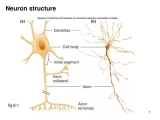
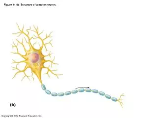
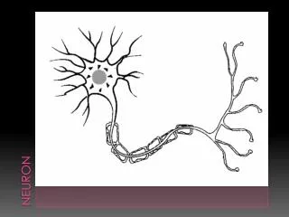
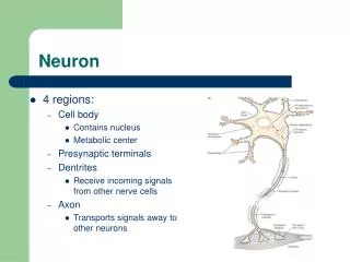
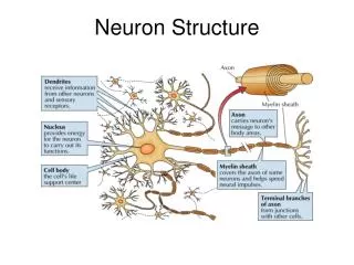
Neuron Structure
Neuron Structure. Synapse. The Synapse. Synthesis of neurotransmitter (NT) Storage and transport of NT within vesicles NT Release Activation of postsynaptic receptors Termination of transmitter effect (e.g. reuptake). Resting Potential.


Neuron Structure
E N D
Presentation Transcript
The Synapse • Synthesis of neurotransmitter (NT) • Storage and transport of NT within vesicles • NT Release • Activation of postsynaptic receptors • Termination of transmitter effect (e.g. reuptake)
Resting Potential Sodium ions are concentrated on the outside of the axon membrane. Potassium ions are concentrated on the inside of the axon membrane. Ion channels are closed. The inside of the axon membrane is more negative that is the outside.
Action Potential • Action potential occurs when the membrane potential rapidly shifts from -70 to +40 mV • Ion channels open in the membrane, allowing sodium ions to enter the axon • Sodium entry shifts the membrane potential toward a positive value • Potential is restored when other channels open, allowing potassium ions to exit the axon
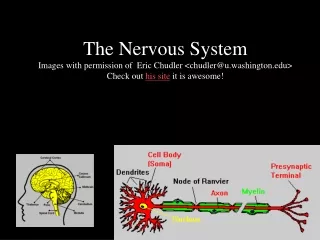
Myelin • Myelin is a fatty, waxy substance coating the axon of some neurons. • Functions: • Speeds neurotransmission • Insulates neurons from each other • Makes neurotransmission more efficient
Neurotransmitters • Serotonin • Acetylcholine • Dopamine • Norepinephrine • Epinephrine • GABA • Endorphins
Brainstem • Brainstem is a primitive portion of brain • Pons: involved in respiration, sleep regulation, dreaming • Medulla: involved in life support functions such as respiration and heart rate • Reticular activating system is an arousal system within the brainstem
Subcortical Brain Areas • Corpus callosum: band of axons that interconnects the hemispheres • Thalamus: sensory relay area • Limbic system: involved in emotionality • Hypothalamus: feeding, fleeing, mating, fighting, homeostasis • Cerebellum: involved in motor control
Cerebral Cortex • Cortexrefers to the outer covering of the brain • Consists of left and right hemispheres • Cortex is divided into lobes • Frontal: Self-awareness, planning, voluntary movement, emotional control, speech, working memory • Parietal: Body sensations • Occipital: Vision • Temporal: Hearing, language comprehension • Localization of function: do discrete circuits carry out different functions?
Nee, et al., STM/LTM article • Damage to Medial Temporal produced LTM deficits while leaving STM in tact. Inferior Temporal = LT visual pattern recognition deficits
Behavioral data confirming STM/LTM distinction • Presentation rate affects primacy but not recency effect • Increased delay affects recency but not primacy effect. (Glanzer & Cunitz, 1966) • Med. Temporal activation for early probes in serial recall paradigm • R infer. parietal for late probes (Talmi, Grady, Goshen-Gottstein, & Moscovitch (2005)
STM/LTM distinction or novelty (MTL) and resistance to distraction (frontal) • Ranganath & Blumenfeld (2005) argue that MTL binds novel items together in single representation. STM storage can be disrupted in patients with MT damage when items are novel (novel items rarely used in most STM studies) • Furthermore patients with frontal damage can perform STM task when distractions are minimized. • Sakai, Rowe, & Passingham (2002), subject did STM spatial task – found greater frontal activity on ‘correct’ trials, less on ‘error’ trials suggesting frontal areas important for filtering distractions. Similar findings for words and pseudo words. • Other evidence suggesting that phonological deficits are found in patients with perisylvian damage, thus this area may not be specifically for STM, but may STM tasks may typically require phonological rehearsal. • STM is ‘attentionally mediated activation’ of LTM representations.
Episodic retrieval and STM retrieval • Similar processes may exist in LTM, STM processes. • Cabeza, et al., 2002 – episodic retrieval: Subs judge probe word as ‘remembered from earlier list,’ ‘known’ or ‘not seen before’. STM: yes/no to probe after study • Same areas of left frontal active in both cases. Anterior frontal may play a monitoring role.