Download
1 / 17
170 likes | 293 Vues
Case 103- 72 y/o man with Chest Pain. History.

E N D
History • Mr. Robertson is a 72-year-old slightly obese male brought to CUMC Emergency Room with a history of severe, unrelenting chest pain. The patient admitted that he has had recurring, crampy chest pain for the past year that is relieved by rest, but he hasn’t seen his physician about it. On Monday night, he felt ‘sick’, but his wife could not convince him to go to the ER. For the next two days, he just didn’t feel right, but he still wouldn’t go. Further questioning reveals that he had unrelenting pressure over his chest since Monday, and he felt like his chest was in a vice. He awoke this morning with severe ‘heartburn’, which seemed to paralyze his left arm and his wife called the rescue squad.
History • PMH (Past Medical History, provided by the patient and Mrs. Ethel Robertson): He has seen his regular physician in the Creighton Medicine Clinic about every year or so. He has had a TURP (transurethral resection of the prostate) for an enlarged prostate 4 years ago. He has been encouraged to lose weight, but he has been unsuccessful in shedding more than a few pounds. There is no history of heart disease and he does not take any cardiac medication. The patient smokes about 1 pack of Camels a day. He has some shortness of breath and a dry cough. • ROS (Review of Systems): Other than as noted above, non-contributory. • FH (Family History): Father died at age 87, mother at age 95. Two brothers still living, ages 70 and 65. One sister died at age 87 of a ‘heart attack’. His four children are still living.
PE • On physical exam, the patient is a mildly obese white male, anxious and in acute distress. An oxygen mask and an IV are in place. He is diaphoretic. Respiratory rate is 24, pulse is 96, blood pressure 135/85 and temperature is 39 degrees Celsius. • HEENT (Head, eyes, ears, nose, and throat): Exam is unremarkable. • Heart: Regular rate and rhythm. No murmurs. • Lungs: Breath sounds are distant and there is dullness to percussion, particularly in the bases. Coarse rales are present in bases bilaterally. Loud rhonchi are heard everywhere. • Abdomen: Obese, but no masses or tenderness noted. • Extremities: Unremarkable. • Rectal: Deferred at this time. • Neurological Exam: Grossly unremarkable.
Develop a problem list. That is, develop a list of medical problems (not diagnoses) that Mr. Robertson has. Which of the problems is/are most important? • From the problem list develop a preliminary differential diagnosis. • What tests and procedures would you order based on the history? What would you do for Mr. Robertson while you are waiting for the lab results?
Hgb - 14.6 g/dL Chemistries: • Hct - 44 % Na - 145 mmol/L • RBC - 4.5 x 10⁶/µL K - 3.0 mmol/L • WBC - 14.6 x 10³/µL Cl - 102 mmol/L • Differential: CO2 - 28 mmol/L • Neutr - 60% - 8.76 x 10³/μL BUN – 28 mg/dL • Bands - 10% - 1.46 x 10³/μL Creatinine - 1.6 mg/dL • Mono - 8% - 1.16 x 10³/μL Glucose - 154 mg/dL • Lymph - 20% - 2.92 x 10³/μL • Eosin - 2% - .29 x 103/μL • Platelets 210x103/µL • UA - Pending • CK, CKMB and Troponin - Pending • EKG - Attached (not read yet) • Chest X-ray - infiltrates in both lungs, most notable in lower lung field
EKG Rate: about 75 minute Rhythm: Generally regular Sinus Rhythm with occasional P.V.C’s. P-R is exactly .2 sec. So we will have to say there is a borderline first degree AV Block. QRS is less than .12 sec. (No B.B.B.). Axis: Left Axis Deviation (nearly -90). No rotation in the horizontal plane. Hypertrophy: Probable left atrial hypertrophy. Left ventricular hypertrophy. Infarction: Significant Q waves in I and AVL. (Coronary ST segments are elevated in I and AVL. ST segments are depressed in vascular V, V2, V3, and V4 status). T waves are flat or inverted in II, III, and AVF and all chest leads. Comment: This patient has a classical acute lateral infarction caused by an occlusion of the Left Circumflex coronary Artery. Coincident with this is a probable occlusion of the Right
An IV is running (D5W) and he has been given, ASA, nitroglycerin and morphine, with some relief of his pain. • After the results of some of the laboratory tests, ECG and chest x-ray, what do you want to do now? What is your differential now? • Predict the results of the lab tests that are pending. If you predict that a test result will be abnormal, why will it be abnormal? What do you think the EKG shows (in very general terms)? • Laboratory results come back with an elevated CK (265 IU/L) with a significant MB fraction (22%), Troponin I of 2.1 ng/mL. • Now that you have the results of these tests, what is your differential diagnosis?
Later that night, Mr. Robertson develops arrhythmias and has a cardiac arrest. After several attempts at resuscitation, a normal cardiac rhythm is attained. Over the next several hours, he is requiring increasing support and is showing signs that major brain damage has occurred. After consultation with the family, he is taken off of life support and dies. An autopsy is performed. A myocardial infarct consistent with 4 days old.There is no evidence of pneumonia, but pulmonary edema is seen.
The autopsy finding are shown. Explain the findings. How will the damaged myocytes differ from normal myocytes? How would you classify this type of necrosis? • Describe the cellular mechanisms occurring in the myocytes at the edge of the infarct. • What caused the myocardial infarct? • What are the risk factors for this process? • If there was no pneumonia, why was there a leukocytosis? • Did this patient have diabetes mellitus? • How would you discuss termination of life support with Mrs. Robertson? In your opinion, what are the major issues?
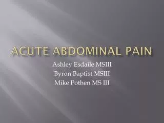
The patient’s heart (weight 630 gm) compared to a normal heart (350 gm)
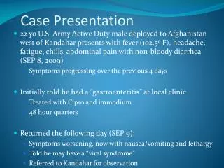
Cross-section of coronary artery - Complete occlusion by a thrombus and severe atherosclerosis.
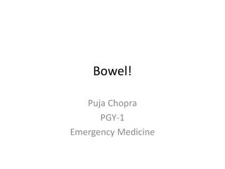
Myocardium from center of infarct - Just outline of cells - pure coagulation necrosis, no inflammatory cells have gotten there yet.
Area from center of infarct, note PMNs Area at hyperemic edge of infarct and grossly normal muscle - The cardiac muscle here is recently ischemic and dying. Note the loss of nuclei, loss of striation, and infiltration by neutrophils