Download
1 / 17
170 likes | 370 Vues
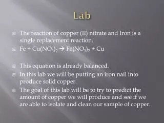
Lab#1 Microscopy & Epithelial Tissue Lama AlAbdi Supervised by: Dr. Reem AlAjmi. Microscope. Genera!y , It is an instrument used to examine and monitor objects we can’t see with the naked eye. It has many applications. Types of microscopes.

E N D
Lab#1 Microscopy & Epithelial Tissue Lama AlAbdi Supervised by: Dr. Reem AlAjmi
Microscope Genera!y, It is an instrument used to examine and monitor objects we can’t see with the naked eye. It has many applications.
Types of microscopes There are different classes and types of microscopes depending on the magnification power, their applications, and how they are operated: Transmission electron microscope (TEM). Scanning electron microscope (SEM). Inverted microscope. Light (compound) microscope.
Compound Microscope The compound microscope is made of 3 systems: The mounting and movement System. The magnification System. The i!umination System.
The body tube: carries the ocular lenses The nose piece: Carries the objective lenses and move them accordingly above the stage The arm: Supports and connects the upper part of the microscope The stage: Horizontal platform upon which the slide of interest rest The coarse focusing knob: for stage movement The fine focusing knob: for image sharpness The base: Supports the microscope • The mounting and movement system
THE MAGNIFICATION SYSTEM The ocular lenses: 5X, 10X, and 15X The objective lenses: Scanning lens: 3.5X or 4X or 5X Low power lens: 10X High power lens: 40X Oil immersion lens: 100X How to calculate the magnification power? Magnification power = ocular lens xobjectivelens. (e.g.) 10X x40X = 400X hint: Don’t forget the unit
The iris diaphragm: controls the amount of light reaching the slide The illuminator: light source The condenser: collects and concentrate the light THE ILLUMINATION SYSTEM
Lab 2ZOO 103 THE EPITHELIAL TISSUE Prepared by : Reem Aldossari
Types of Epithelial Tissue • Epithelial tissue can be divided into two groups depending on the number of layers of which it is composes. stratified epithelium: which consist of more than one layer simple epithelium: which consist of only one layer squamous epithelium columnar epithelium cuboidal epithelium ( Squamous,cuboidal,columnar) Skin of Toad ( Frog ) Intestine Collecting tubule Stomach Bowman’s capsule Side view Thyroid gland Lining of Mouth Top view
Types of Epithelial Tissue • Epithelial tissue can be divided into two groups depending on their function of which it is composes. • Neuro_epithium Glandular epithelium • Covering “surface” epithelium. • epithelium of cells specialized to produce • secretion. • (Goblet cell) • The Simple. • Stratified.
2 1 • Glandular Epithelium
Stratified sequamous epithelium in skin keratinized and non keratinized