Download
1 / 55
550 likes | 705 Vues
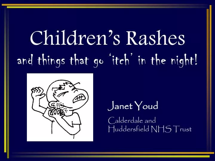
Children’s Rashes and things that go ‘itch’ in the night!. Janet Youd Calderdale and Huddersfield NHS Trust. Objectives. To understand the terminology used in describing rashes and skin lesions. To illustrate some common rashes seen in children. Background.

E N D
Children’s Rashesand things that go ‘itch’ in the night! Janet Youd Calderdale and Huddersfield NHS Trust
Objectives • To understand the terminology used in describing rashes and skin lesions. • To illustrate some common rashes seen in children.
Background • Ill children often present with several symptoms, one of the most common being a rash. • Any attempt to identify a rash should come after the systematic assessment of a sick child.
SYSTEMATIC APPROACH TO RASH IDENTIFICATION • History/Examination • Distribution (Body Location) • Morphology of primary and secondary lesions. • Configuration / Arrangement • Pattern of Distribution • Consult Textbook
History • Associated symptoms, timings and sequence of onset. Aggravating/relieving factors • Recent contacts/symptoms in family members/peers • Social history/pets • Recent travel • Immunisation history • Past medical history • Drug history • Known allergies
Examination • Ensure privacy • Suitable environment • Will need full systems examination if signs of systemic illness • Look: • Total skin evaluation (including folds) • Evaluate hair and nails • Feel: • Subtle changes in texture
Distribution • Scattered/Generalised: spread throughout the body • Localised: involve only a selected part
MACULE • Derived from the Latin for Stain. • Used to describe changes in colour or consistency without elevation above the surface of the surrounding skin. • Typically less than 1cm • e.g. Freckles
PATCH As a macule but greater than 1cm. • e.g. Vitiligo or Café au Lait spot
PAPULE Raised, palpable skin lesions smaller than 1cm in diameter that may or may not have a different colour from the surrounding skin.
NODULE • As a papule but greater than 1cm.
PLAQUE Raised, palpable skin lesion greater than 1cm in diameter. Usually confined to the superficial dermis. • Typically seen in psoriasis.
WHEALS Raised circumscribed, oedematous plaques that usually are pink or pale and tend to be present only temporarily.
VESICLE A raised lesion of less than 1cm that contains clear serous fluid. • Typical of herpes simplex.
BULLAE As a vesicle but greater than 1cm. It may be superficial within the epidermis or may be situated in the dermis below. • Commonly Seen in partial Thickness burns.
PUSTULES Papules filled with pus. • Commonly seen in patients with acne.
PURPURA General name for the escape of red blood cells into the skin. Petechiae are less than 0.5cm
Secondary Lesions • Excoriations • Scratch marks
Secondary Lesions • Lichenification • Typical thickening of the skin. Often seen in patients with chronic pruritus.
Secondary Lesions • Crusts • Raised lesions produced by dried serum and blood cell remnants.
Secondary Lesions • Erosions • Depressed lesions produced whenever the epidermis is either removed or sloughed. They are moist, usually red and well circumscibed. Classically seen in chicken pox after rupture of a vesicle.
Secondary Lesions • Ulcers • Depressed lesions produced whenever not only the epidermis but also part of (or all of) the dermis is gone.
Secondary Lesions • Fissures • Depressed lesions that present as narrow and linear skin cracks. They penetrate through the epidermis and reach at least part of the dermis.
Terms to describe configuration • Annular: Ring shaped
Terms to describe configuration • Linear: Lesions arranged in a line
Terms to describe configuration • Reticular: Net-like clusters
Pattern of distribution • Clustered: Grouped • Confluent: Multiple lesions that blend together • Dermatomal: Distributed along neurocutaneous dermatomes
Information • 1-2 day history of general malaise and low grade pyrexia. • Initially noticed itchy, scattered rash of discrete lesions of varying morphology. Some are macular papular, that develop to vesicles. • Within 24 hours developed some secondary crusts, whilst new lesions continued to erupt over then next 4-5 days. • There are some ulcers within the mouth.
Information • 3 day history of high fever, cough, red and watery eyes. Child miserable. • Developed non-itchy, scattered, maculopapular confluent rash. Started at the hairline and worked down. • Koplick spots are noted on buccal mucosa.
Information • Tiny pink macules starting on face and working down the body, associated with low grade pyrexia and slight post-auricular lymphadenopathy. Rash fades quickly.
Information • Systemically well child with discrete papules (1-5 mm) with a central dimple, clustered and localised to chest and abdomen.
Information • Tingling skin sensation followed by clustered or isolated vesicles, localised to specific area, commonly face/lips. Develop secondary crusts. Resolve 5-14 days.
Information • Localised flaccid blisters rupture and form ‘golden’ crusts. Spreading occurs readily. Most commonly seen around the nose and mouth.
Information • Rapid onset (hours) flu-like symptoms. May have scattered non-itchy maculopapular rash followed by development of petechiae and purpura.
Information • Child presents with non-itchy purpuric rash localised to legs and buttocks. May also have haematuria +/- abdominal pain. He is otherwise well.
Information • Sudden onset widespread wheals following ingestion of strawberrries.
Information • Localised very itchy oedematous and erythematous lesion may develop to vesicles followed by secondary crusting and scaling.
Information • Intensely itchy, localised papules and vesicles, some ‘burrows’ may be seen. Often secondary excoriation noted. Commonly found between fingers and on flexor surfaces at elbows, knees and groins.