Dementia
Dementia . So what does it look like?. So what do you think???.


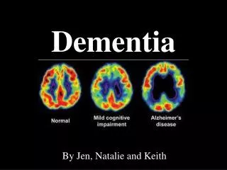
Dementia
E N D
Presentation Transcript
Dementia So what does it look like?
So what do you think??? 65 yo male retired attorney presents with change in behavior over the past 3mths. Pt. has been increasingly vulgar in language, passes gas in public, and has abandoned all dinner table etiquette. Pt was once very subdued and refined.
Pick’s Disease • Pick's disease is a rare neurodegenerative disorder, occurring 1/10 as often as Alzheimer's disease. • Personalityproblems are more common in Pick's disease, while memory problems are more common in Alzheimer's disease. • Frontal and temporal lobe atrophy, with sparing of the occipital and parietal lobes. The cerebral gyral atrophy in Pick's disease is so severe that some of the gyri are extremely thin which is also known as knife blade atrophy. • PET scan shows decreased activity in the frontotemporal region.
So who’s my next victim? • 48 yo female is brought in by her daughter. She reports a one week history of her mother forgetting how to cook her signature dish, disoriented to time, forgetting important appointments, and trouble reading her mail. PMH significant for DM, HTN, CRI. Family history significant for CAD, Alzheimer’s, DM, and HTN.
That’s right!!! Vascular Dementia • Two magnetic resonance imaging proton-density transaxial cuts from the brain of a hypertensive patient with vascular dementia. Note extensive involvement of white matter, which appears as hyperintense signals. • Vascular dementia can result from ischemic or hemorrhagic brain damage. • The three most common mechanisms: single, strategically placed infarcts; multiple cortical infarcts; and subcortical small-vessel disease.
There’s been an accident! • You are in the ER and an ambulance comes in with a 78 yo lady who has fallen on the sidewalk of a residential neighborhood. Residents of the neighborhood noticed the lady walking earlier but no one recognized her which is strange because the community is very small and everyone knows everyone else. The lady seems confused and somewhat belligerent when attempting to interview her. She has a bump on her forehead on PE. CT scan is ordered. What is the cause of her condition?
Alzheimer’s Disease • CT shows diffuse cerebral atrophy with widened sulci and dilatation of the ventricles .
Alzheimer’s Disease • Patients with AD most commonly present with progressive memoryloss, to which other spheres of cognitive impairment are added over several years. • After memory loss occurs, patients may also have language disorders (eg, anomia, progressive aphasia) and impairment in their visuospatial skills and executive functions. • The anatomic pathology of AD includes neurofibrillary tangles; senile plaques at the microscopic level; and cerebrocortical atrophy, which predominantly involves the association regions and particularly the medial aspect of the temporal lobe.
Alzheimer’s Disease • Atrophy caused by Alzheimer's Disease processes can be visualized using structural imaging techniques such as T1-weighted MRI scans. Areas such as the medial temporal lobe (outlined in white) are not severely affected in normal aging (A - from a 74-year-old healthy female) and are especially affected in AD (B - from a 72-year-old female with moderate AD).
Can you solve this mystery? • 24 yo male is brought to the ER by friends because he has demonstrated over the course of several months reduced work productivity, poor concentration, mental slowness, decreased libido, and forgetfulness. However, they are really concerned because for that last few days he is experiencing increasing clumsiness, difficulty speaking, and memory loss. Pt is unable to communicate effectively in the interview and his friends stated that all they knew about his medical history is that he has some “problem with his immune system” but he has never discussed it entirely with them. Image 1 – CT and Image 2 – MRI.
HIV-1 Encephalopathy and AIDS Dementia • CT scan of the brain of a patient with AIDS dementia complex (ADC) shows diffuse atrophy and ventricular enlargement and attenuation of periventricular white matter (these lesions represent areas of infarction and demyelination). • T2-weighted MRI shows ventricular enlargement and large areas of hyperintense signal in the subcortical white matter of both frontal lobes.