Download
1 / 49
490 likes | 820 Vues
Amino acids, peptides, and proteins. IUG, Fall 2018 Dr Tarek Zaida. Amino Acids, Peptides, and Proteins. Proteins are naturally occurring polymers composed of amino acid units joined one to another by amide (or peptide) bonds.

E N D
Amino acids, peptides, and proteins IUG, Fall 2018 Dr TarekZaida
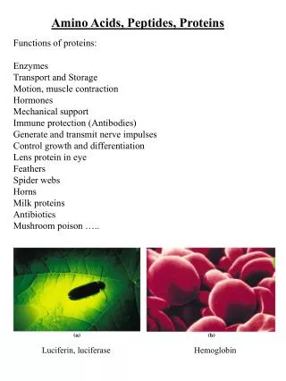
Amino Acids, Peptides, and Proteins • Proteins are naturally occurring polymers composed of amino acid units joined one to another by amide (or peptide) bonds. • Spider webs, animal hair and muscle, egg whites, and hemoglobin (the molecule that transports oxygen in the body to where it is needed) are all proteins.
Peptides • Are oligomers of amino acids that play important roles in many biological processes. • For example, the peptide hormone insulin controls our blood sugar levels, • Thus, proteins, peptides, and amino acids are essential to the structure, function, and reproduction of living organisms.
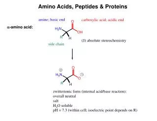
Amino Acids carboxyl group • The amino acids obtained from protein hydrolysis are α-amino acids. • That is, the amino group is on the α-carbon atom, the one adjacent to the carboxyl group. amino group • a-carbon is chiral (except for glycine) • at pH 7.0 uncharged amino acids are zwitterions • amino acids have a tetrahedral structure a-carbon side chain
With the exception of glycine, where R is H, α-amino acids have a stereogenic center at the α-carbon. • All except glycine are therefore optically active. • They have the L configuration relative to glyceraldehyde. • Note that the Fischer projection, used with carbohydrates, is also applied to amino acids
The following table lists the 20 α-amino acids commonly found in proteins. • The amino acids are known by common names. • Each also has a three-letter abbreviation based on this name, which is used when writing the formulas of peptides, and a one-letter abbreviation used to describe the amino acid sequence in a protein. • The amino acids in the following table are grouped to emphasize structural similarities.
Of the 20 amino acids listed in the table, 12 can be synthesized in the body. • The other 8, those with names shown in a blue color and referred to as essential amino acids, cannot be synthesized by adult humans and therefore must be included in the diet in the form of proteins.
The Amphoteric Nature of Amino Acids • Amino acids are amphoteric. • They can behave as acids and donate a proton to a strong base, or • they can behave as bases and accept a proton from a strong acid. • These behaviors are expressed in the following equilibria for an amino acid with one amino and one carboxyl group:
The isoelectric Point • If placed in an electric field, the amino acid will therefore migrate toward the cathode (negative electrode) at low pH • and toward the anode (positive electrode) at high pH. • At some intermediate pH, called the isoelectric point (pI), the amino acid will have a net charge of zero. • It will be unable to move toward either electrode. • Each of the amino acids has a characteristic isoelectric points
Peptides • Amino acids are linked in peptides and proteins by an amide bond between the carboxyl group of one amino acid and the α-amino group of another amino acid. • Emil Fischer, who first proposed this structure, called this amide bond a peptide bond. • A molecule containing only two amino acids joined in this way is a dipeptide:
Write out the abbreviated formulas for all possible tripeptide isomers of: 1. Leu—Ala—Met 2. Gly—Ala—Ser
A spider’s Web A spider’s web is a device built by the spider to trap prey. Spider silk, a protein, is the main component of the web. Silk is composed largely of β-sheets, a fundamental unit of protein structure. Many proteins have β-sheets; silk is unique in being composed almost entirely of β-sheets.
Protein Three-Dimensional Structurecomposed of primary, secondary, and tertiary structures • Functioning proteins are not simply long polymers of amino acids. • These polymers fold to form discrete three-dimensional structures with specific biochemical functions. • The amino acid sequence is called the primary structure. • Three-dimensional structure resulting from a regular pattern of hydrogen bonds between the NH and the CO components of the amino acids in the polypeptide chain is called secondary structure.
The three-dimensional structure becomes more complex when the R groups of amino acids far apart in the primary structure bond with one another. • This level of structure is called tertiary structure and is the highest level of structure that an individual polypeptide can attain. • However, many proteins require more than one chain to function. • Such proteins display quaternary structure, which can be as simple as a functional protein consisting of two identical polypeptide chains or as complex as one consisting of dozens of different poly-peptide chains.
1. The Primary Structure of Proteins • The number and sequence of the amino acids in the protein chain. • Primary structure is stabilized by peptide bonds. • A slight change in the amino acid sequence (replacement of an amino acid with another), may change the entire protein.
2. Secondary Structure • Polypeptide chains can fold into regular structures. • Two types of secondary structure elements: 1. α-helix 2. β-sheet
1. α-helix • Is a coiled structure stabilized by intrachain hydrogen bonds. • Each turn of the helix contains a bout 3.6 amino acids. • Hair & wool are examples of protein with helical structure, both contain keratin.
A ribbon depiction shows the –carbon atoms and side chains (green). (B) A side view of a ball-and-stick version depicts the hydrogen bonds (dashed lines) between NH and CO groups. (C) An end view shows the coiled backbone as the inside of the helix and the side chains (green) projecting outward. (D) A space-filling view of part C shows the tightly packed interior core of the helix.
β-sheet • Beta sheets are stabilized by hydrogen bonding between polypeptide strands instead of a single polypeptide strand, • The β-sheet is composed of two or more polypeptide chains called β-strands. • A β-strand is almost fully extended rather than being tightly coiled as in the α-helix. • The distance between adjacent amino acids along a β-strand is approximately 3.5 Å, in contrast with a distance of 1.5 Å along an α-helix. • The side chains of adjacent amino acids point in opposite directions.
Examples of proteins with β-sheet are protein in silk (fibroin), and proteins that bind fatty acids The structure of a β-strand. The side chains (green) are alternatively above and below the plane of the strand. The bar shows the distance between two residues
Tertiary Structure • The specific folding and bending of the coils into specific layers or fibers. • This level of structure is the result of inter-actions between the R groups of the peptide chain. • It is the tertiary structure that gives proteins their biological activity. • Tertiary structure is stabilized by several types of bonds.
Types of Bonds that stabilize tertiary structure • Hydrogen bonds • Disulfide bridges • Hydrophobic interactions • Salt bridges between positively & negatively charged groups within the protein. • Nonpolar amino acids are folded on the inside of the protein, and polar amino acids are on the outside where they react with water to form polar group interactions.
Quaternary Structure • It occurs when a protein has at least 2 units combine together to form a complex.
Percent Composition • Average percentage of nitrogen in protein is 16%. • 16% Nitrogen = 1/6 of protein content. • Protein is the major food containing nitrogen. • Chemists can determine the amount of protein present in food substance by deter-mining the amount of nitrogen present. • Calculation of protein in food can be done by multiplying the weight of nitrogen by 6 and converting it to a percentage of the total.
% protein in food = grams of Nitrogen x 6 • Example: • If 100 g of food yield 4 g of nitrogen on chemical analysis. Calculate the % protein in the sample? • Since the amount of nitrogen in protein is 1/6 of the total amount of protein, • Then the amount of protein present in food is: 4g Nitrogen x 6 = 24 24 g protein in 100 g sample food; meaning 24%
Classification of Proteins • Proteins can be classified into three classes: 1. Simple protein 2. Conjugated proteins 3. Derived proteins
Simple proteins: they give amino acids or their derivative (polypeptides) upon hydrolysis • Conjugated proteins: they give amino acids and other compounds (non protein compound) upon hydrolysis • Derived proteins: Are produced from simple or conjugated proteins upon chemical action or enzyme on proteins. As derived proteins one can consider proteoses, peptones, polypeptides, tripeptides, and dipeptides.
Classification Of Proteins According To Solubility • Simple proteins are classified according to their solubility in various solvents and also as to whether they are coagulated by heat.
Classification Of Proteins According To Composition • Conjugated proteins can be classified into different types: • Nucleoproteins: (nucleic acid/ chromosomes) • Glycoproteins: (carbohydrates/ mucin in saliva) • Phosphoproteins: (Phosphate/ casein in milk) • Chromoproteins: (Chromophore/ hemoglubin, cytochrome) • Lipoproteins: (lipids/ fibrin in blood) • Metalloproteins: Metals/ ceruloplasmin
Classification Of Proteins According To Function According to their biological function, proteins can be classified into various classes: 1. Structural proteins: Collagen (in connective tissues), keratin (in hair). 2. Contractile proteins: Myosin, actin (muscle contaction) 3. Storage proteins: Ferritin (storage of iron) 4. Transport proteins:Hemoglobin (transfers oxygen) 5.Hormones: Insulin ( metabolism of carbohydrates) 6. Enzymes: Pepsin (digestion of proteins) 7. Protective proteins: gamma-globulin (antibody formation) 8. Toxin: Venoms (poisons)
Classification Of Proteins According To Shape 1. Globular proteins: • Folded into a shape of ball. • Are soluble in water or form colloidal disper-sions. • Examples: Hemoglobin, Albumin, globulins 2. Fibrous proteins • Consists of parallel polypeptide chains that are coiled and stretch out. • Are insoluble in water. • Examples: collagen, fibrin, myosin.
Properties of Proteins • Proteins have two characteristic properties: • Colloidal Nature • Denaturation
1. Colloidal Nature • When proteins are in water, they form a colloidal dispersion. • Due to this property, proteins will not pass through a membrane. • This is very important because proteins present in bloodstream can not pass through the capillaries & should remain in blood-stream. • Therefore there should be no protein in the urine. • So if there is a protein in urine, it indicates that there is a damage in the kidney membranes-possibly nephritis.
2. Denaturation • Unfolding & rearrangement of secondary and tertiary structure of a protein without breaking the peptide bonds. • A denatured protein loses its activity • If denaturing conditions are mild, protein will restore their active structure if these conditions of denaturing are reversed. • If denaturation is drastic, the process is irreversible; the protein will coagulate or precipitate from solution.
Factors or Reagents Causing Denaturation 1. Alcohol - It causes irreversible denaturation. - 70% alcohol is used to disinfect bacteria because of its ability to coagulate proteins. - Alcohols form hydrogen bonds, that compete with original hydrogen bonds
2. Salts of heavy metals HgCl2 (mercuric chloride) and AgNO3 (silver chloride) cause irreversible denaturation by disrupting salt bridges & disulfide bridges. 3. Heat • If heat is gentle, protein will denature reversibly. • If heat is vigorous, protein will denature irreversibly by disrupting several types of bonds.
Examples: 1.Egg-white coagulates on heating. 2. In bacteria heat destroys & coagulates proteins, hence in hospitals, the sterilization of instruments and clothing for use, need high temperature. 3. Determination of proteins present in urine can be done by heating a sample of urine which will cause the coagulation of any protein present.
4. Alkaloidal Reagents (tannic acid & picric acid) - Both form insoluble compounds with proteins. They denature proteins irreversibly by dis-rupting salt bridges and hydrogen bonds. 5. Radiation UV & X-ray cause coagulation of proteins. They denature proteins irreversibly by disrupting the hydrogen bonds and hydropho-bic bonds.
pH Changing the pH will disrupt hydrogen bonds and salt bridges causing irreversible denaturing. (H2SO4, HCl, HNO3). 7. Oxidizing & reducing agents - Bleach (chlorine), nitric acid, both oxidizing agents. - SO32- (sulfites) & oxalates are reducing agents. Both denature proteins by disrupting disulfide bridges.
8. Salting out: • The vast majority of proteins are insoluble in saturated salt solutions and precipitate out unchanged. • It is possible to separate proteins from other proteins in a mixture by placing the mixture in a saturated solution of (NH4)2SO4, Na2SO4 or NaCl. • The protein is precipitated out and removed by filtration.