Download
1 / 72
770 likes | 893 Vues
Learn about the vital functions and operations of the endocrine system, including hormonal actions, target cell specificity, and control mechanisms. Explore the role of hormones and their effects on metabolic activities and growth regulation.

E N D
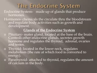
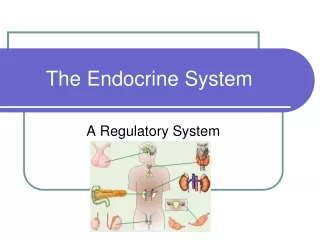
Endocrine System Functions • Maintenance of the internal environment in the body (maintaining the optimum biochemical environment). • Influences metabolic activities • Integration and regulation of growth and development. • Control, maintenance and instigation of sexual reproduction
Endocrine vs Nervous System • Nervous system performs short term crisis management • Endocrine system regulates long term ongoing metabolic • Endocrine communication is carried out by endocrine cells releasing hormones • Alter metabolic activities of tissues and organs • Target cells
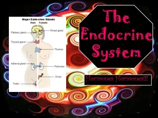
The Endocrine System Figure 18.1
Endocrine System: Overview • Endocrine glands – pituitary, thyroid, parathyroid, adrenal, pineal, and thymus • The hypothalamus has both neural functions and releases hormones • The pancreas and gonads produce both hormones and exocrine products • Other tissues and organs that produce hormones –, cells of the small intestine, stomach, kidneys, and heart
Hormones • Hormones – chemical substances secreted by cells into the extracellular fluids • Regulate the metabolic function of other cells • Have lag times ranging from seconds to hours • Tend to have prolonged effects • Are classified as amino acid-based hormones, or steroids • Eicosanoids – biologically active lipids with local hormone–like activity
Mechanism of Hormone Action • Hormones produce one or more of the following cellular changes in target cells • Alter plasma membrane permeability • Stimulate protein synthesis • Activate or deactivate enzyme systems • Induce secretory activity • Stimulate mitosis
Types of Hormones • Amino acid based – most hormones belong to this class, including: • Amines, thyroxine, peptide, and protein hormones • Steroids – gonadal and adrenocortical hormones • Eicosanoids – leukotrienes and prostaglandins
Classification of Hormones Figure 18.2
Hormone Action • Hormones stimulate target cell activity by one of two mechanisms • Second messengers involving: • Regulatory G proteins • Amino acid–based hormones • Direct gene activation involving steroid hormones • The precise response depends on the type of the target cell
AA-Based Hormone Action: PIP-Calcium Figure 16.2b
G Proteins and Hormone Activity Figure 18.3
Steroid Hormones • Diffuse into target cells to bind and activate a specific intracellular receptor • Hormone-receptor complex travels to the nucleus and binds a DNA-associated receptor protein prompting DNA transcription and protein synthesis
Steroid Hormones Figure 16..3
Target Cell Specificity • Hormones circulate to all tissues but only activate target cells • Target cells must have specific receptors to which the hormone binds • These receptors may be intracellular or located on the plasma membrane • Examples of hormone activity • ACTH receptors are only found on certain cells of the adrenal cortex • Thyroxin receptors are found on nearly all cells of the body
Target Cell Activation • Target cell activation depends on three factors • Blood levels of the hormone • Relative number of receptors on the target cell • The affinity of those receptors for the hormone • Up-regulation – target cells form more receptors in response to the hormone • Down-regulation – target cells lose receptors in response to the hormone
Interaction of Hormones • Three types of hormone interaction • Permissiveness – one hormone cannot exert its effects without another hormone being present • Synergism – more than one hormone produces the same effects on a target cell • Antagonism – one or more hormones opposes the action of another hormone
Hormone Concentrations in the Blood • Hormones circulate in the blood in two forms – free or bound • Steroids and thyroid hormone are attached to plasma proteins • All others are unencumbered • Concentrations of circulating hormone reflect: • Rate of release • Speed of inactivation and removal from the body • Hormones are removed from the blood by: • Degrading enzymes • The kidneys • Liver enzyme systems
Control of Hormone Release • Blood levels of hormones: • Are controlled by negative feedback systems • Vary only within a narrow desirable range • Hormones are synthesized and released in response to humoral, neural, and hormonal stimuli
Humoral Stimuli • Secretion of hormones in direct response to changing blood levels of ions and nutrients • Example: concentration of calcium ions in the blood • Declining blood Ca2+ concentration stimulates the parathyroid glands to secrete PTH (parathyroid hormone) • PTH causes Ca2+ concentrations to rise and the stimulus is removed
Hormonal Stimuli • Release of hormones in response to hormones produced by other endocrine organs • The hypothalamic hormones stimulate the anterior pituitary • In turn, pituitary hormones stimulate targets to secrete still more hormones
Endocrine Control: Three Levels of Integration • Hypothalamic stimulation–from CNS • Pituitary stimulation–from hypothalamic trophic hormones • Endocrine gland stimulation–from pituitary trophic hormones
Feedback control of Endocrine Secretion Figure 18.8a
Neural Stimuli • ANS efferent nerve fibers stimulate hormone release • Preganglionic sympathetic nervous system (SNS) fibers stimulate the adrenal medulla to secrete catecholamines Figure 16.4b
Nervous System Modulation • The nervous system modifies the stimulation of endocrine glands and their negative feedback mechanisms • The nervous system can override normal endocrine controls Ex. Control of blood glucose levels: Normally the endocrine system maintains blood glucose. Under stress, the body needs more glucose The hypothalamus and the sympathetic nervous system are activated to supply ample glucose
Endocrine Glands Figure 18.1
Major Endocrine Organs: Pituitary (Hypophysis) • The hypothalamus sends a chemical stimulus to the anterior pituitary to releasing hormones stimulate the synthesis and release of hormones • TRH - Thyrotropin releasing hormone (TRH) >> release of TSH • CRH - Corticotropin releasing hormone (CRH) >> release of ACTH • GnRH - Gonadotropin releasing hormone (GNRH) >> release of FSH and LH • Pituitary gland – two-lobed organ that secretes nine major hormones • Neurohypophysis – posterior lobe (neural tissue) - receives, stores, and releases hormones from the hypothalamus • Adenohypophysis – anterior lobe, made up of glandular tissue - synthesizes and secretes a number of hormones
Pituitary (Hypophysis) • Releases nine important peptide hormones • All nine bind to membrane receptors and use cyclic AMP as a second messenger Figure 16.5
Pituitary (Hypophysis) • Nuclei of the hypothalamus synthesize oxytocin and antidiuretic hormone (ADH) • These hormones are transported to the posterior pituitary • The six hormones of the adenohypophysis: • Are abbreviated as GH, TSH, ACTH, FSH, LH, and PRL • Regulate the activity of other endocrine glands Figure 18.7
Hormones of the Adenohypophysis • Growth hormone (GH or somatotropin) • Thyroid stimulating hormone (TSH) • Adrenocorticotropic hormone (ACTH) • Follicle stimulating hormone (FSH) • Luteinizing hormone (LH) • Prolactin (PH or PRL)
Growth Hormone (GH) • Produced by somatotropic cells of the anterior lobe that: • Stimulate most cells, but target bone and skeletal muscle • Promote protein synthesis and encourage the use of fats for fuel • Most effects are mediated indirectly by somatomedins • GH stimulates liver, skeletal muscle, bone, and cartilage to produce insulin-like growth factors • Direct action promotes lipolysis and inhibits glucose uptake
Growth Hormone (GH) • Antagonistic hypothalamic hormones regulate GH • Growth hormone–releasing hormone (GHRH) stimulates GH release • Growth hormone–inhibiting hormone (GHIH) inhibits GH release
Metabolic Action of Growth Hormone Figure 16.6
Thyroid Stimulating Hormone • Tropic hormone that stimulates the normal development and secretory activity of the thyroid gland • Triggered by hypothalamic peptide thyrotropin-releasing hormone (TRH) • Rising blood levels of thyroid hormones act on the pituitary and hypothalamus to block the release of TSH
Adrenocorticotropic Hormone • Stimulates the adrenal cortex to release corticosteroids • Triggered by hypothalamic corticotropin-releasing hormone (CRH) in a daily rhythm • Internal and external factors such as fever, hypoglycemia, and stressors can trigger the release of CRH
Gonadotropins • Gonadotropins – follicle-stimulating hormone (FSH) and luteinizing hormone (LH) • Regulate the function of the ovaries and testes • FSH stimulates gamete (egg or sperm) production • Absent from the blood in prepubertal boys and girls • Triggered by the hypothalamic gonadotropin-releasing hormone (GnRH) during and after puberty
Functions of Gonadotropins • In females • LH works with FSH to cause maturation of the ovarian follicle • LH works alone to trigger ovulation (expulsion of the egg from the follicle) • LH promotes synthesis and release of estrogens and progesterone • In males • LH stimulates interstitial cells of the testes to produce testosterone • LH is also referred to as interstitial cell-stimulating hormone (ICSH)
Prolactin • In females, stimulates milk production by the breasts • Triggered by the hypothalamic prolactin-releasing hormone (PRH) • Inhibited by prolactin-inhibiting hormone (PIH) • Blood levels rise toward the end of pregnancy • Suckling stimulates PRH release and encourages continued milk production
Neurohormones: secreted into the Blood by Neurons Figure 7-12: Synthesis, storage, and release of posterior pituitary hormones
Posterior Lobe of the Pituitary Gland (Neurohypophysis) • Contains axons of hypothalamic nerves • Neurons of the supraoptic nucleus manufacture antidiuretic hormone (ADH) aka vasopressin • Decreases the amount of water lost at the kidneys • Elevates blood pressure • Neurons of the paraventricular nucleus manufacture oxytocin • Stimulates contractile cells in mammary glands • Stimulates smooth muscle cells in uterus
Oxytocin • Oxytocin is a strong stimulant of uterine contraction • Regulated by a positive feedback mechanism to oxytocin in the blood • This leads to increased intensity of uterine contractions, ending in birth • Oxytocin triggers milk ejection (“letdown” reflex) in women producing milk • Synthetic and natural oxytocic drugs are used to induce or hasten labor • Plays a role in sexual arousal and satisfaction in males and non-lactating females
Antidiuretic Hormone (ADH) • ADH helps to avoid dehydration or water overload - prevents urine formation • Osmoreceptors monitor the solute concentration of the blood • With high solutes, ADH is synthesized and released, thus preserving water • With low solutes, ADH is not released, thus causing water loss from the body • Alcohol inhibits ADH release and causes copious urine output
Thyroid Gland • The largest endocrine gland composed of colloid filled follicles • Colloid = thyroglobulin + iodine fills the lumen of the follicles and is the precursor of thyroid hormone • Thyroid hormones end up attached to thyroid binding globulins (TBG) produced by the liver , some are attached albumin • Other endocrine cells, the parafollicular cells, produce the hormone calcitonin
Synthesis of Thyroid Hormone Figure 16.8
Thyroid hormones • Involves of two closely related iodine-containing compounds • Both are non-steroid hormones • Triiodothyronine (T3) has two tyrosines with three bound iodine atoms • Thyroxine (T4) has two tyrosine molecules plus four bound iodine atoms • T4 and T3 bind to thyroxine-binding globulins (TBGs) • Both bind to target receptors, but T3 is ten times more active than T4 • Mechanisms of activity are similar to steroids
Thyroid Hormones • Regulates metabolism • Promotes glycolysis, gluconeogenesis, glucose uptake • Glucose oxidation • Increasing metabolic rate • Heat production • Regulates growth and development • Regulates tissue growth • Increases protein synthesis • Developing skeletal and nervous systems • Maturation and reproductive capabilities • C Cells produce Calcitonin - helps regulate calcium concentration in body fluids • Helps maintaining blood pressure
Thyroid hormones • Bound to mitochondria, thereby increasing ATP production • Bound to receptors activating genes that control energy utilization • Exert a calorigenic effect