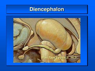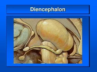
Diencephalon and telencephalon
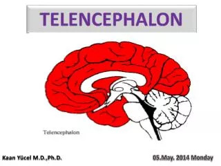
Diencephalon and telencephalon. Surface structure. medial surface of the diencephalon interthalamic adhesion or massa intermedia connects two thalami bundle of nerve fibers called stria medullaris thalami dorsal surface: concealed by fornix, curves over the thalamus. Components.

Diencephalon and telencephalon
E N D
Presentation Transcript
Surface structure • medial surface of the diencephalon • interthalamic adhesion or massa intermedia connects two thalami • bundle of nerve fibers called stria medullaris thalami • dorsal surface: • concealed by fornix, curves over the thalamus
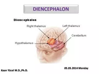
Components • Diencephalon • Thalamus, the largest component. Receives sensory information and involves emotion, memory, sleep • Subthalamus, ventral to thalamus. reticular formation, red nucleus, and substantia nigra project fibers into subthalamus • Epithalamus, dorsalmedial to thalamus. Includes pineal body • Hypothalamus, outgrow into posterior pituitary, controls endocrine system
Thalamus • Almost all the nuclei send fibers to the cortex. However, there is no interconnection fibers among different nuclei groups within the thalamus.
Thalamus • Reticular nucleus • not connected with the brain stem, unknown function • Intralaminar nuclei: • Receive afferent fibers from reticular formation of the brain stem. • Also receive collateral fibers from spinothalamic and trigeminothalamic fibers (main destination is the ventral posterior nuclei of thalamus), also receives fibers from cerebellum, and the globus pallidus. There is extensive connection between intralaminar nuclei and frontal, parietal lobes. Connection between reticular formation to cortex through this area involves alertness and consciousness.
Ventral group of Nuclei • Medial geniculate body: • Afferent fibers: • inferior brachium: fibers from inferior colliculus, which receive lateral lemniscus. Medial geniculate body receives fibers from both sides but predominately from contralateral side. The bilateral distribution is caused by crossed fiber from ventral cochlear nucleus and by the commissural fibers between the inferior colliculi. • Efferent: • ipsilateral side of the temporal cortex, conscious awareness of sound, and the adjacent association area is responsible for hearing recognition with previous experience.
Ventral group of Nuclei • Lateral geniculate body: • has six layer of neurons (crossed fibers to 1, 4, 6 and uncrossed fibers to 2, 3, 5) • Afferent fibers: • fibers from optic nerve. Point to point project from retina to lateral geniculate body. • Efferent fibers: • geniculocalcarine tract: ipsilateral side of the occipital cortex
Ventral posterior nucleus • pathway for conscious awareness of somatic sensation, like pain, temperature info, touch, proprioception • Afferent fibers: • all fibers of medial lemniscus, and most of the spinothalamic and trigeminothalamic tracts • also fibers from vestibular nuclear complex and fibers from gustatory nucleus concerning with sensation about position, and taste, respectively. • There is a detailed projection of the opposite body on the ventral posterior nucleus. The lower limb is represented in its dorsolateral part with upper limb in an intermediate position and the head the most medial. The importance (not area) of certain body part determines the area in the ventral posterior nucleus. Same as the cerebral cortex. • Ventral posterior nucleus also receives afferent fibers involving taste, and vestibular information • The medial region of the nucleus that receives info from head is also referred as ventral posteromedial division (VPm), the lateral portion for the rest of the body called ventral posterolateral division (VPl). • Efferent fibers: • project to the somesthetic cortex of parietal lobe.
Ventral lateral nucleus • Includes posterior and anterior divisions. • Afferent fibers: • From cerebellum • Efferent fibers: • Enter internal capsule and proceed to frontal lobe
Other nuclei • Posterior group of nucleus • Associated with pain reception • Lateral group of nuclei: • lateral dorsal nucleus: part of the limbic system • lateral posterior nucleus: connected with the somatosensory associate ion area of the parietal lobe • Medial group of nuclei: • mediodorsal nucleus: may be related with memory, seen with Korsakoff's syndrome, and perception of pain • Afferent fibers are from entorhinal cortex, amygdaloid body and corpus striatum • medioventral nucleus: little is known • Anterior group of nuclei: • part of the limbic system
Thalamic syndrome • most often caused by vascular lesion, usually in ventral posterior region • Opposite side of the body: • proprioception and the sensations of touch, pain, and temperature are affected contralaterally • could also involve emotional disturbance
Subthalamus • sensory fasciculi: • includes medial lemniscus, spinothalamic tract, and trigeminothalamic tract • substantia nigra and red nucleus extend from midbrain into subthalamus • Efferent fiber from pallidus pallidus (part of the basal ganglia, more later) contained in two bundles, lenticular fasciculus and ansa lenticularis. Both terminate in the subthalamus.
Epithalamus • The epithalamus consists of the hebenular nuclei and the pineal gland. • Habenular neuclei • Afferent fibers are received through the stria medularis thalami • Efferent fibers known as habenulointerpeduncular fasciculus terminate in the interpeduncular nuclei in the roof of the interpeduncular fossa. The interpeduncular nuclei influence neurons in the hypothalamus and preganglionic autonomic neurons via relays in reticular formation.
Pineal gland • influenced by light, regulate time and day night changes and onset of puberty in human
Hypothalamus • The hypothalamus can be divided into medial and lateral zones
Medial Zone • contains suprachiasmatic, tuberal, and mamillary regions. • Within suprachiasmatic region, it contains supraoptic, paraventricular, suprachiasmatic and anterior nuclei • Supraoptic: large cells, above the optic chiasm • Paraventricular nucleus: large cells • Both supraoptic and paraventricular nuclei secrete hormones (ADH and Oxytoxin), form the hypothalamo-hypophysial tract • Suprachiasmatic neurons are releasing action potential spontaneously at rugular rhythm and pattern. Axons from retina leave the optic chisma and termiate into this area and regulate the light-dark cycle. • Anterior nucleus: some neurons in this area are twice as many in males as in females (one possible reason is females experience neuronal death after 4 yr. old) • Tuberal region contains ventromedial, dorsomedial, and infundibular nuclei • Mamillary region contains mamillary body and the posterior nucleus.
Lateral Zone large nerve cells sparsely located and collectively constitute the lateral nucleus of the hypothalamus. • Lateral zone includes the lateral tuberal nucleus • Fibers of hypothalamus • hypothalamus served as the main integrator of the autonomic and endocrine systems
Afferent fibers • Ascending afferents: convey information of visceral origin • Most of the ascending fibers originate from nuclei of reticular formation • Medial forebrain bundle originates from septal area, with fibers originate from intermediate and lateral olfactory areas, conducts information related to basic emotional drives and the sense of smell, smaller in humans • Fibers from amygdaloid body also involve smell and emotional drives by stria terminalis (arise from amygdaloid body and ends in the preoptic area, anterior nucleus, and the septal area). • Fornix: • originates from hippocampal formation (hippocampus) and parahippocampal gyrusends in the hypothalamus
Efferent fibers • Originate from hypothalamus (paraventricular nucleus), starts as periventricular fibers underneath the third ventricle and continue into the dorsal longitudinal fasciculus in the periaqueductal gray matter of the midbrain. Some terminate into dorsal nucleus of vagus nerve and other may end in the intermediolateral cell column and sacral autonomic nucleus (via spinal cord). Thus the influence from hypothalamus to the preganglionic fibers of sympathetic and parasympathetic nervous system. • Hypothalamus also influence cells in motor nuclei of trigeminal, facial nerves, nucleus of ambiguus and the hypoglossal nucleus to regulate muscles used in feeding and drinking. Motor neurons in spinal cord is also affected by hypothalamus thus in shivering response. • Fibers originate from mamillary body • mamillothalamic fasciculus: project to anterior nuclei of the thalamus • collateral branches of the fibers of the mamillothalamic fasciculus are also called mamillotegmental fasciculus, end in nuclei of the reticular formation of the midbrain and pons.
Functional aspect of hypothalamus • Stimulation of anterior hypothalamus( preoptic area and anterior nucleus) • slowing heart rate, vasodilation, lowering blood pressure, salivation, increased peristalsis in the gastrointestinal gract, contraction of urinary bladder, sweating. Action of parasypathtic system. • Stimulation of posterior and lateral nuclei: • sympathetic activation: cardia accelaeration, elevation of blood pressure, cessation of peristalsis, dalation off pupil, hyperglycemia. • Hypothalamus is also responsible for regulating temperature control, Neurons in anterior hypothalamus are sensitive to temperature changes. If temperature is high, heat loss mechanism will be activated including cutaneous vasodilation, sweating. Lesion of anterior hypothalamus = = > hyperthemia.
Functional aspect of hypothalamus • Cells in posterior hypothalamus are responsible for lowering blood temperature, triggering heat conservation response, including coetaneous vasoconstriction, and shivering. Lesion in posterior hypothalamus impairs temperature regulation, causes hypothemia. • Hypothalamus is also responsible for regulation food intake and water intake. Hunger or feeding center is located in the lateral zone and satiety center in the region of ventromedial nucleus. Lesion of ventromedial nucleus results excessive food intake and obesity. • Hypothalamus and pituitary • Hypothalamus constitute the posterior part of pituitary. Secrete ADH and Oxytoxin.