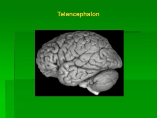
TELENCEPHALON
TELENCEPHALON. Kaan Yücel M.D.,Ph.D. 05.May. 201 4 Monday. 1. Telencephalon is composed of… . Cerebral hemispheres Sulci , fissures , gyri , lobes

TELENCEPHALON
E N D
Presentation Transcript
TELENCEPHALON Kaan Yücel M.D.,Ph.D. 05.May. 2014Monday
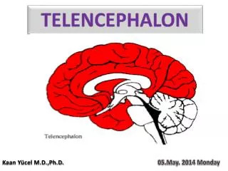
1. Telencephalon is composed of… Cerebralhemispheres Sulci, fissures, gyri, lobes partially separated by a deep longitudinal fissure, and which fill the area of the skull above the tentorium cerebelli and are subdivided into lobes based on their position. Basalganglia Insula Ventricularsystem Twolateralventricles, thirdventricle, fourthventricle White mattertracts Commissural, association, andprojectionfibres
2. Sulci & Gyri & Lobes Precentralgyrus
8. Brodmannareas Neocortex6 layers Paleocortex3-5 layers Archicortex3 layers 1909 Cytoarchitecture 47 functionalareas
8. Brodmannareas 4 8 3 5 7 6 9 1 10 46 39 46 19 10 2 4 4 4 5 43 41, 42 18 47 22 17 21 11 37 38
11. Fourthventricle FrontalLobe. ParietalLobe. OccipitalLobe. SeptumPellucidum. a. Rostrum of CorpusCallosum.b. Body of CorpusCallosum.c. Splenium of CorpusCallosum.10. Pons. 11.Medulla Oblongata. 12. Cerebellum. 13. SpinalCord. 14. Fourthventricle. 15. SinusConfluence.
12. Basalganglia Distributedset of brain structures in the telencephalon, diencephalon, and mesencephalon. The forebrain structures include : Caudate nucleus Putamen Nucleusaccumbens(or ventral striatum) Globuspallidus Corpus striatum
12. Basalganglia Caudatenucleus C-shaped structure closely associated with the lateral wall of the lateral ventricle. largest at its anterior pole (the head), and its size diminishes posteriorly as it follows the course of the lateral ventricle (the body) all the way to the temporal lobe (the tail), where it terminates at the amygdaloid nuclei.
12. Basalganglia Putamen- Caudateseparatedbyanterior limb of the internal capsule Connectedbybridges of cellsacrosstheinternalcapsule A striatedlook striatumorneostriatum Caudate+ Putamen=Striatum= Main recepient of afferentinput. Globuspallidus= Majorefferentoutputleavesfrom.
12. Basalganglia Third ventricle MRI of thebrain, T1-weighted axialcut.1, Putamen. 2, Pallidum. 3, Caudatenucleus. 4, Insula. 5, Lateralventricle. 6, Thalamus.
FunctionalAnatomy of theBasalGanglia Traditionally, the basal ganglia have been viewed as motor structures. It is only in the past 20 years that it has been recognized that these structures also may have a role in cognition and emotion.
FunctionalAnatomy of theBasalGanglia Alexander and his colleagues that the motor circuit is involved in ‘‘thecontrol of movementdirectionand in thescaling of movementamplitudeorvelocity’’ and ‘‘in theprogrammingandinitiation of internallygeneratedmovements.’’
FunctionalAnatomy of theBasalGanglia EXTRAPYRAMIDAL SYSTEM • GABA, dopamine, acetylcholine, and glutamine • Multiplepathways in thebasalgangliabothwith both excitatory and inhibitory functions. • Input to the basal ganglia is received from both thecerebral cortex and the thalamus. • Lesions in the basal ganglia result in uncoordinated and disorganized movement.
12. Basalganglia MRI of thebrain, T1-weighted coronalcut.1, Lateral ventricle. 2, Caudate nucleus. 3, Putamen. 4, Temporal lobe (left side). 5, Sylvian fissure (lateral sulcus).
13. Commissuralfibers CORPUS CALLOSUM Freitag CM, Luders E, Hulst HE, Narr KL, Thompson PM, Toga AW, Krick C, Konrad C. Total brain volume and corpus callosum size in medication-naïve adolescents and young adults with autism spectrum disorder.Biol Psychiatry. 2009;66(4):316-319. Kitayama N, Brummer M, Hertz L, Quinn S, Kim Y, Bremner JD.Morphologic alterations in the corpus callosum in abuse-related posttraumatic stress disorder: a preliminary study.J NervMent Dis. 2007;195(12):1027-1209. Ballmaier M, Kumar A, Elderkin-Thompson V, Narr KL, Luders E, Thompson PM, Hojatkashani C, Pham D, Heinz A, Toga AW. Mapping callosal morphology in early- and late-onset elderly depression: an index of distinct changes in cortical connectivity. Neuropsychopharmacology. 2008;33(7):1528-1536. Black SE, Moffat SD, Yu DC, Parker J, Stanchev P, Bronskill M. Callosal atrophy correlates with temporal lobe volume and mental status in Alzheimer's disease.Can J Neurol Sci. 2000;27(3):204-209. VenkatasubramanianG, Jayakumar PN, Reddy VV, Reddy US, Gangadhar BN, Keshavan MS Corpuscallosumdeficits in antipsychotic-naïveschizophrenia: evidenceforneurodevelopmentalpathogenesis.PsychiatryRes. 2010;182(2):141-145.
Genu of corpuscallosum Forcepsminor Anteriorlimb of internalcapsule Septumpellucidum Caudatenucleus Putamen Globuspallidus Posteriorlimb of internalcapsule Thalamus Splenium of corpuscallosum Forcepsmajor
13. Commissuralfibers ANTERIOR COMMISSURE POSTERIOR COMMISSURE
15. Projectionfibers 1st neuron 2nd neuron 3rd neuron Internalcapsule Anteriorlimb [3] Posteriorlimb [8] Genu
Sensoryhomunculus Motor homunculus
Sensoryhomunculus Motor homunculus
The cerebral arterial circle (of Willis) is formed at the base of the brain by the interconnecting vertebrobasilar and internal carotid systems of vessels. • 1anterior communicating artery connecting the left and right anterior cerebral arteries to each other; • 2posterior communicating arteries, one on each side, connecting the internal carotid artery with the posterior cerebral artery.