Osteoporosis
Osteoporosis is a condition characterized by reduced bone strength, increasing fracture risk, especially in post-menopausal women. Defined by a T-score lower than -2.5, it affects over 10 million Americans. Key risk factors include age, gender, smoking, low body weight, and certain medications. Diagnosis involves DXA scans, with treatment options such as bisphosphonates, SERMs, and hormone therapy. Lifestyle modifications and monitoring are crucial for those at risk. Awareness and early intervention are vital for effective management of osteoporosis.

Osteoporosis
E N D
Presentation Transcript
Osteoporosis 06/25/12 José L. González, PGY3
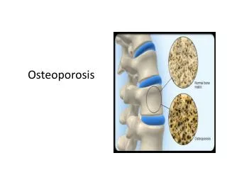
Definition • Reduction in bone strength increase risk of fx • T-score: < -2.5 SDs • T-score: 30 yo, matched for sex and race • Osteopenia: <-1 to 2.5 SDs
Epidemiology • >10 million • 8 million women & 2 million men • Most fractures occur in women w/ osteopenia • Rate of collesfx increases initially, later hip • May be due to the way we fall • Vertebral > Hip > Colles
Risk Factors • Age, female sex, cigarette smoking, prior fxs, low body weight, excess etoh • Meds: glucocorticoids, cyclosporine, heparin, levothyroxine, anticonvulsants • Diseases • Vision • Dementia • Chronic inflammatory diseases • RA • Crohns
Bone Remodeling • Bone mass is 50-80% heritable • Peak skeletal mass early adulthood. Constant mass 30-45 yoa, then increased resorption • Estrogens, androgens, vitamin D, PTH • 2 functions • Repair microdamage of the skeleton • Maintain [Ca2+] serum
Risk Factors: Parathyroid Hormone • Kidneys • 1. ↑ hydroxylation 1,25OH vit D • 2. decreased Ca2+ loss • Small Intestine • ↑Ca2+ absorbtion • Bone • Release of Ca2+
Risk Factors: vitamin D / Calcium • Calcium: RDI 1000 – 1200 • Vitamin D: RDI 800-1000 units daily • RFs for low vit D: • High latitude • Low intake • Chronic liver or renal disease • Estrogen • Physical Activity • ↓risk in rural communities
Diagnosis • US, CT scan, single energy absorptiometry, DXA • DXA • Lumbar and hip m.c. used • Z-score: age matched • T-score: 30 yo, race and sex matched
Who to test: • All women > 65 yoa • Estrogen deficient women @ risk • Vertebral abnormality of x-ray suggestive • Primary hyper parathyroidism • Steroids > 7.5mg x 3 months • Monitoring response to meds • Repeat @ 2 year intervals
Who to treat: • T-score < -2.5 SDs • Post-menopausal women w/ RFs • RFs: age, prior fx, family hx, low weight, smoking, RA, etoh • FRAX calculator
Treatment: SERMs, PTH, Calcitonin • Raloxifene: • tx & pv of ER+ breast ca • Tx & pv of osteoporosis • PTH: in small amounts • Calcitonin: (intranasal) • Hormone produced by thyroid • decreases osteoclast activity • Decreases vert. fxs only
Treatment: Estrogens • Estrogens (in the form of combined OCPs) • Decrease fracture risk by 50% • Increase risk of MI by 29% • Increase stroke risk by 40% dementia 2x • Increase risk of breast ca by 26% • Decrease risk of colon ca by 37% • Increase risk of VTE by 100% • 10,000 patients: • prevents (5 hip, 6 colles & 44 clinical fxs) • leads to (8 breast cas, 8 MIs, 18 VTEs)
Treatment: Bisphosphonates • Alendronate: • 5mg x 2 yrs, 10mg x 9 months 90% ↓vert. fx, 50% ↓hip fx • 70mg PO dose once weekly • Risedronate: • use in steroid-induced OP • Ibandronate • Zolendronic Acid: • 70mg IV q yearly • for hyperCa2+
Bisphosphonates: Side Effects • Osteonecrosis of the jaw • hypocalcemia • GI side effects • Esophagitis, ulceration • Contraindicated in strictured esophagus • Drink w/ full glass H20 and remain upright x 30min
Sources: • Harrison’s Principles of Internal Medicine. 18th ed. New York, NY: McGraw-Hill; 2005:946.