Download
1 / 155
1.64k likes | 2.14k Vues
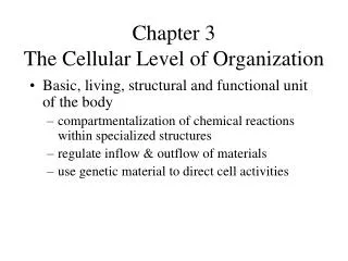
3 The Cellular Level of Organization. An Introduction to Cells. Cell Theory Developed from Robert Hooke’s research Cells are the building blocks of all plants and animals All cells come from the division of preexisting cells Cells are the smallest units that perform all vital

E N D
3 The Cellular Level of Organization
An Introduction to Cells • Cell Theory • Developed from Robert Hooke’s research • Cells are the building blocks of all plants and animals • All cells come from the division of preexisting cells • Cells are the smallest units that perform all vital physiological functions • Each cell maintains homeostasis at the cellular level
An Introduction to Cells • Sex Cells (Germ Cells) • Reproductive cells • Male sperm • Female oocyte (a cell that develops into an egg) • Somatic Cells • Soma = body • All body cells except sex cells
Figure 3-1 Anatomy of a Model Cell Plasma membrane Nonmembranous organelles Membranous organelles Secretory vesicles Centrosome and Centrioles Cytoplasm contains two centrioles at right angles; each centriole iscomposed of 9 microtubule triplets in a 9 0 array CYTOSOL FunctionsEssential formovement ofchromosomesduring cell division;organization ofmicrotubules incytoskeleton Centrosome Centrioles
Figure 3-1 Anatomy of a Model Cell Plasma membrane Nonmembranous organelles Cytoskeleton Membranous organelles Proteins organized in fine filaments orslender tubes Microfilament FunctionsStrength andsupport; movement ofcellular structuresand materials Microtubule Plasma Membrane Lipid bilayer containing phospholipids, steroids, proteins, and carbohydrates Freeribosomes FunctionsIsolation;protection; sensitivity; support;controls entryand exit ofmaterials Cytosol (distributesmaterials by diffusion)
Figure 3-1 Anatomy of a Model Cell Microvilli Membrane extensionscontaining microfilaments FunctionIncrease surfacearea to facilitateabsorption of extra-cellular materials Plasma membrane Nonmembranous organelles Membranous organelles
Figure 3-1 Anatomy of a Model Cell Cilia Cilia are long extensionscontaining microtubuledoublets in a 9 2 array (notshown in the model cell) Plasma membrane FunctionMovement of material over cell surface Nonmembranous organelles Membranous organelles Proteasomes Hollow cylinders of proteolyticenzymes with regulatory proteins at their ends FunctionsBreakdown and recycling of damaged or abnormal intracellular proteins Ribosomes RNA proteins; fixed ribosomesbound to rough endoplasmicreticulum, free ribosomesscattered in cytoplasm FunctionProtein synthesis
Figure 3-1 Anatomy of a Model Cell Golgi apparatus Stacks of flattened membranes(cisternae) containing chambers FunctionsStorage, alteration, and packaging of secretory products and lysosomal enzymes Mitochondria Double membrane, with innermembrane folds (cristae)enclosing important metabolicenzymes FunctionsProduce 95% of the ATPrequired by the cell Endoplasmic reticulum (ER) Network of membranouschannels extendingthroughout the cytoplasm Rough ERmodifies andpackages newlysynthesized proteins NUCLEUS FunctionsSynthesis of secretoryproducts; intracellularstorage and transport Smooth ERsynthesizes lipids and carbohydrates Peroxisomes Vesicles containingdegradative enzymes FunctionsCatabolism of fats and otherorganic compounds,neutralization of toxiccompounds generated inthe process Plasma membrane Nonmembranous organelles Membranous organelles
Figure 3-1 Anatomy of a Model Cell Plasma membrane Nonmembranous organelles Membranous organelles Peroxisomes Vesicles containingdegradative enzymes FunctionsCatabolism of fats and otherorganic compounds,neutralization of toxic compounds generated in the process Freeribosomes Lysosomes Vesicles containingdigestive enzymes FunctionsIntracellular removal ofdamaged organelles orpathogens
Figure 3-1 Anatomy of a Model Cell Chromatin NUCLEUS Nuclearenvelope Nucleoplasm containingnucleotides, enzymes,nucleoproteins, andchromatin; surrounded by a double membrane,the nuclear envelope NUCLEOPLASM Nucleolus(site of rRNAsynthesis andassembly ofribosomalsubunits) Nuclearpore Functions:Control of metabolism; storage and processing of genetic information;control of proteinsynthesis
3-1 Plasma Membrane • Extracellular Fluid (Interstitial Fluid) • A watery medium that surrounds a cell • Plasma membrane (cell membrane) separatescytoplasm from the extracellular fluid • Cytoplasm • Cytosol = liquid • Intracellular structures collectively known as organelles
3-1 Plasma Membrane • Functions of the Plasma Membrane • Physical Isolation • Barrier • Regulation of Exchange with the Environment • Ions and nutrients enter • Wastes eliminated and cellular products released
3-1 Plasma Membrane • Functions of the Plasma Membrane • Sensitivity to the Environment • Extracellular fluid composition • Chemical signals • Structural Support • Anchors cells and tissues
3-1 Plasma Membrane • Membrane Lipids • Phospholipidbilayer • Hydrophilic heads — toward watery environment, both sides • Hydrophobic fatty-acid tails — inside membrane • Barrier to ions and water — soluble compounds
3-1 Plasma Membrane • Membrane Proteins • Integral Proteins • Within the membrane • Peripheral Proteins • Bound to inner or outer surface of the membrane
3-1 Plasma Membrane • Membrane Proteins • Anchoring Proteins (stabilizers) • Attach to inside or outside structures • Recognition Proteins (identifiers) • Label cells as normal or abnormal • Enzymes • Catalyze reactions
3-1 Plasma Membrane • Membrane Proteins • Receptor Proteins • Bind and respond to ligands (ions, hormones) • Carrier Proteins • Transport specific solutes through membrane • Channels • Regulate water flow and solutes through membrane
3-1 Plasma Membrane • Membrane Carbohydrates • Proteoglycans, glycoproteins, and glycolipids • Extend outside cell membrane • Form sticky “sugar coat” (glycocalyx) • Functions of the glycocalyx • Lubrication and Protection • Anchoring and Locomotion • Specificity in Binding (receptors) • Recognition (immune response)
Figure 3-2 The Plasma Membrane EXTRACELLULAR FLUID Phospholipidbilayer Integral proteinwith channel Integralglycoproteins Glycolipidsof glycocalyx Hydrophobictails Plasmamembrane Cholesterol Hydrophilicheads Peripheralproteins Gatedchannel Cytoskeleton(Microfilaments) 2 nm CYTOPLASM
3-2 Organelles and the Cytoplasm • Cytoplasm • All materials inside the cell and outside the nucleus • Cytosol (intracellular fluid) • Dissolved materials • Nutrients, ions, proteins, and waste products • High potassium/low sodium • High protein • High carbohydrate/low amino acid and fat • Organelles • Structures with specific functions
3-2 Organelles and the Cytoplasm • The Organelles • Nonmembranous organelles • No membrane • Direct contact with cytosol • Include the cytoskeleton, microvilli, centrioles, cilia, ribosomes, and proteasomes • Membranous organelles • Covered with plasma membrane • Isolated from cytosol • Include the endoplasmic reticulum (ER), the Golgi apparatus, lysosomes, peroxisomes, and mitochondria
3-2 Organelles and the Cytoplasm Nonmembranous Organelles Six types of nonmembranous organelles Cytoskeleton Microvilli Centrioles Cilia Ribosomes Proteasomes
3-2 Organelles and the Cytoplasm • The Cytoskeleton • Structural proteins for shape and strength • Microfilaments • Intermediate filaments • Microtubules
3-2 Organelles and the Cytoplasm • The Cytoskeleton • Microfilaments — thin filaments composed of the protein actin • Provide additional mechanical strength • Interact with proteins for consistency • Pair with thick filaments of myosin for muscle movement
3-2 Organelles and the Cytoplasm • The Cytoskeleton • Intermediate filaments — mid-sized between microfilaments and thick filaments • Durable (collagen) • Strengthen cell and maintain shape • Stabilize organelles • Stabilize cell position
3-2 Organelles and the Cytoplasm • The Cytoskeleton • Microtubules — large, hollow tubes of tubulin protein • Attach to centrosome • Strengthen cell and anchor organelles • Change cell shape • Move vesicles within cell (kinesin and dynein) • Form spindle apparatus
3-2 Organelles and the Cytoplasm • The Cytoskeleton • Thick filaments • Myosin protein in muscle cells
Figure 3-3a The Cytoskeleton Microvillus Microfilaments Plasma membrane Terminal web Mitochondrion Intermediatefilaments Endoplasmicreticulum Microtubule Secretoryvesicle The cytoskeleton provides strength andstructural support for the cell and its organelles. Interactions between cytoskeletal components are also important in moving organelles and in changing the shape of the cell.
Figure 3-3b The Cytoskeleton Microvillus Microfilaments Terminal web The microfilaments andmicrovilli of an intestinal cell.Such an image, produced by a scanning electron microscope, is called a scanning electron micrograph (SEM) (SEM 30,000).
Figure 3-3c The Cytoskeleton Microtubules (yellow) in a living cell, as seen afterspecial fluorescent labeling(LM 3200).
3-2 Organelles and the Cytoplasm • Microvilli • Increase surface area for absorption • Attach to cytoskeleton • Centrioles in the Centrosome • Centrioles form spindle apparatus during cell division • Centrosome cytoplasm surrounding centriole • Cilia • Small hair-like extensions • Cilia move fluids across the cell surface
Figure 3-4a Centrioles and Cilia Microtubules Centriole. A centriole consistsof nine microtubule triplets(known as a 9 0 array). A pairof centrioles orientated at rightangles to one another occupiesthe centrosome. Thismicrograph, produced by a transmission electronmicroscope, is called a TEM.
Figure 3-4b Centrioles and Cilia Plasma membrane Microtubules Basal body Cilium. A cilium contains nine pairs ofmicrotubules surrounding a central pair(9 2 array). The basal body to which the cilium is anchored has a structure similar to that of a centriole.
Figure 3-4c Centrioles and Cilia Return stroke Power stroke Ciliary movement. Action of a single cilium. During the power stroke, the cilium isrelatively stiff; during the return stroke, itbends and returns to its original position.
3-2 Organelles and the Cytoplasm • Ribosomes • Build polypeptides in protein synthesis • Two types • Free ribosomes in cytoplasm • Manufacture proteins for cell • Fixed ribosomes attached to ER • Manufacture proteins for secretion • Proteasomes • Contain enzymes (proteases) • Disassemble damaged proteins for recycling
3-2 Organelles and the Cytoplasm • Membranous Organelles • Five types of membranous organelles • Endoplasmic reticulum (ER) • Golgi apparatus • Lysosomes • Peroxisomes • Mitochondria
3-2 Organelles and the Cytoplasm • Endoplasmic Reticulum (ER) • Endo- = within, plasm = cytoplasm, reticulum = network • Cisternae are storage chambers within membranes • Functions • Synthesis of proteins, carbohydrates, and lipids • Storage of synthesized molecules and materials • Transport of materials within the ER • Detoxification of drugs or toxins
3-2 Organelles and the Cytoplasm • Endoplasmic Reticulum (ER) • Smooth endoplasmic reticulum (SER) • No ribosomes attached • Synthesizes lipids and carbohydrates • Phospholipids and cholesterol (membranes) • Steroid hormones (reproductive system) • Glycerides (storage in liver and fat cells) • Glycogen (storage in muscles)
3-2 Organelles and the Cytoplasm • Endoplasmic Reticulum (ER) • Rough endoplasmic reticulum (RER) • Surface covered with ribosomes • Active in protein and glycoprotein synthesis • Folds polypeptide protein structures • Encloses products in transport vesicles
Figure 3-5a The Endoplasmic Reticulum Nucleus Rough endoplasmicreticulum with fixed(attached) ribosomes Smoothendoplasmicreticulum Ribosomes The three-dimensional relationships between the rough and smooth endoplasmic reticula are shown here. Cisternae
Figure 3-5b The Endoplasmic Reticulum Rough endoplasmicreticulum with fixed(attached) ribosomes Freeribosomes Smoothendoplasmicreticulum TEM 111,000 EndoplasmicReticulum Rough endoplasmicreticulum and freeribosomes in thecytoplasm of a cell.
3-2 Organelles and the Cytoplasm • Golgi Apparatus • Vesicles enter forming face and exit maturing face • Functions • Modifies and packages secretions • Hormones or enzymes • Released through exocytosis • Renews or modifies the plasma membrane • Packages special enzymes within vesicles for use in the cytoplasm
Figure 3-6a The Golgi Apparatus Secretoryvesicles Secretoryproduct Transportvesicles Here is a three-dimensionalview of the Golgi apparatuswith a cut edge.
Figure 3-6b The Golgi Apparatus Golgi apparatus TEM 42,000 This is a sectional view of the Golgiapparatus of an active secretory cell.
Figure 3-7 Protein Synthesis Protein releasedinto cytoplasm Smooth ER Ribosome DNA Rough ER mRNA Cytoplasm Nucleus Transportvesicle Nuclearpore
Figure 3-7 Protein Synthesis Cisternae Lysosome Exocytosis atcell surface Secretingvesicle Cis faceof Golgicomplex Trans faceof Golgicomplex Membranerenewalvesicle Membranerenewal
Figure 3-7 Protein Synthesis DNA mRNA Cytoplasm Nucleus
Figure 3-7 Protein Synthesis Ribosome Rough ER mRNA Cytoplasm
Figure 3-7 Protein Synthesis Protein releasedinto cytoplasm Ribosome
Figure 3-7 Protein Synthesis Rough ER Cytoplasm