
Rheumatoid arthritis
E N D
Presentation Transcript
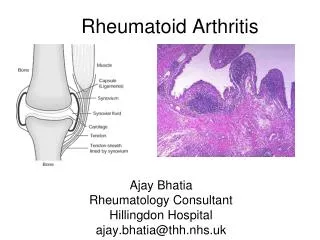
Rheumatoid arthritis • Chronic systemic inflammatory disorder that may affect many tissues and organs – skin , blood vessels , heart ,lungs and muscles – but principally attacks the joints , producing a non suppurative proliferative and inflammatory synovitis that often progresses to destruction of the articular cartilage and ankylosis ( stiffness ) of the joints .
Diagnostic criteria for rheumatoid arthritis Four of the following seven criteria must be met: • Morning stiffness for minimum 1 hour everyday , at least for 6 weeks • Soft tissue swelling (arthritis) of three or more joints present for at least 6 weeks • Swelling (arthritis) of the proximal interphalangeal, metacarpophalangeal, or wrist joints present for at least 6 weeks • Symmetrical swelling present for at least 6 weeks • Subcutaneous nodules. • Positive test for rheumatoid factor. • Radiographic features of RA (erosions and/or periarticular osteopenia in hand and/or wrist joints )
Risk factors • Cause is multifactorial in people with genetic susceptibility • HLA DR4 and DR1 are associated, especially in severe disease • Possible infective aetiology, although no organism has been demonstrated • Onset is more common in winter
Pathogenesis • Believed that RA is an autoimmune disease triggered by exposure of a genetically susceptible host to an unknown arthritogenic antigen. Autoimmune reaction Activation of CD4 +T cells and lymphocytes and the release of inflammatory mediators and cytokines Destroy the joints
Cont .. • Pathogenesis of the disease depends upon : • The nature of the autoimmune reaction • The mediators of tissue injury • Genetic susceptibility • The arthritogenic antigen (s)
The nature of the autoimmune reaction • Activated CD4+ Tcells and B lymphocytes Produce Cytokines Increase immune complex deposition Joint destruction
The mediators of tissue injury : cytokines play an important role stimulates Produce by macrophages and synovial lining cells that are activated by the T – cells in the joints Cytokines ( TNF and IL-1) Synovial cells Activates Produce Osteoclasts Various mediators of inflammation (PG ) and Matrix metalloprotineases (cartilage destruction) Promotes Bone destruction Progressive joint damage
Synovium ( hyperplastic ) Rich in inflammatory cells becomes adherent to and grows over the articular surface , forming a Pannus - an inflammatory exdudate overlying the synovial cells on the inside of a joint stimulates Release of IL-1 , platelet – derived growth factor Prostaglandin Cause cartilage destruction and bone erosion
Cont .. • Genetic susceptibility ( HLA) • Arthritogenic antigen : • No firm evidence has definitively identified a microbial organism as an etiologic agent
Clinical course • Begins slowly and insidiously Initially : • Malaise • Fatigue • Generalised musculoskeletal pain after which the joints become clearly involved
Patterns of joints involvement: • Small joints ----------- large joints Hands • MCP • PIP Elbows Wrist Feets Ankle Knees MTPIP Lumbosacral regions Hip (less) Cervical (usually )
Cont … Involved joints : • Swollen • Warm • Painful • Particularly stiff on arising or following inactivity
Symptoms • Starts as an insidious symmetrical polyarthritis, often with non-specific systemic symptoms : - • Pain • Swelling • Stiffness, especially early morning or after inactivity • Fatigue, fever and weight loss are common
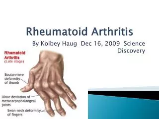
Signs Classically: • Symmetrical, distal, small joint arthritis involving proximal interphalangeal, metacarpophalangeal, wrists, metatarsophalangeal, ankles, knees and cervical spine joints. • Hips are less commonly affected. Hand deformities : - • Ulnar deviation (due to rupture of the collateral ligaments at the MCP joints ) • Swan neck deformity ( due to rupture of the volar(both the palm and sole) plate of the PIP joints ) and • Boutonniere's deformity (due to rupture of central extensor )of the fingers . • Muscle wasting and tendon rupture.
Systemic involvement Eyes: • scleritis and episcleritis. Skin: • Leg ulcers ,rashes, nail fold infarcts. Rheumatoid nodules: • Common; may occur in eyes, subcutaneous, lung, heart and occasionally vocal cords. Neurological: • peripheral nerve entrapment, atlanto-axial subluxation, polyneuropathy,
Cont .. Respiratory system: • pleural involvement, pulmonary fibrosis, bronchiolitis, Cardiovascular system: • pericardial involvement, valvulitis and myocardial fibrosis, Kidneys: • nephropathy, amyloidosis. Liver: • mild hepatomegaly and abnormal transaminases common. Others: • thyroid disorders, osteoporosis, depression, splenomegaly.
Differential Diagnosis Includes: • Viral arthritis • Connective-tissue disease (e.g. systemic lupus erythematosus) • Polyarticular gout • Osteoarthritis • Septic arthritis (particularly if monoarthritis) • Medical conditions presenting with arthropathy, e.g. sarcoidosis , thyroid disease, infective endocarditis, multiple myeloma
Diagnosis • Diagnosis is essentially clinical and there is no diagnostic investigation. Investigations are important in assessment and exclusion of other possible diagnoses. • ESR, CRP : usually raised but may be normal. • Full blood count: • normochromic, normocytic anaemia and reactive thrombocytosis common in active disease. Raised ferritin but low serum iron concentration and total iron binding capacity. • Liver function tests: mild elevation of alkaline phosphatase • Uric acid/synovial fluid analysis: excludes polyarticular gout. • Urinalysis: microscopic haematuria/proteinuria may suggest connective tissue disease.
Cont .. Rheumatoid Factor: • positive in 60-70% of patients (and 5% of normal population). • An antibody to a substance called cyclic citrullinated peptide (CCP) has been found to be more specific than Rheumatoid Factor in rheumatoid arthritis and may be more sensitive in erosive disease. Antinuclear antibody: • positive in SLE and related conditions; also in up to 30% of rheumatoid arthritis patients
Radiology: • Show soft tissue swelling, • periarticular osteopenia, • loss of joint space, • erosions • Deformity • Radial deviation of the wrist • Ulnar deviation of the fingers • Swan neck deformity ( flexon – hyperextension deformity ).
Treatment • Drugs used to treat RA are: Nonsteroidal Anti-Inflammatory Drugs (NSAIDs) – • used to reduce inflammation and relieve pain • e.g : - aspirin, ibuprofen, indomethacin and COX-2 inhibitors such as valdecoxib and celecoxib. Analgesic Drugs – • Used to relieve pain, but don’t necessarily have an effect on inflammation. • e .g : - acetaminophen, propoxyphene, mepeidine and morphine. Glucocorticoids or Prednisone – • prescribed in low maintenance doses to slow joint damage caused by inflammation. Disease Modifying Antirheumatic Drugs (DMARDs) – • used with NSAIDs and/or prednisone to slow joint destruction caused by RA over time. • Examples : - methotrexate, injectable gold, penicillamine, azathioprine, chloroquine, hydroxychloroquine, sulfasalazine and oral gold.
Cont.. Biologic Response Modifiers – • directly modify the immune system by inhibiting proteins called cytokines, which contribute to inflammation. • Examples abatacept, etanercept, infliximab, adaliumumab and anakinra. Protein-A Immuonoadsorption Therapy (dialysis-like technique) • This is not a drug, but a therapy that filters blood to remove antibodies and immune complexes that promote inflammation. DMARDs, particularly methotrexate, have been the standard for aggressively treating RA. • Recently, studies have shown that the most aggressive treatment for controlling RA may be the combination of methotrexate and another drug, particularly biologic response modifiers. • The dual drug treatment seems to create a more effective treatment, especially for people who may not have success with or who have built up a resistance to, methotrexate or another drug alone. • Combination with methotrexate: lefluonomide (Arava), etanercept (Enbrel), adalimumab (Humira) and infliximab (Remicade
Surgery • done to improving the quality of life with RA. Synovectomy – • When one or two joints are affected more severely than others, this procedure is used to reduce the amount of inflammatory tissue by removing the diseased synovium or lining of the joint. • It may result in less swelling and pain and the slowing or prevention of further joint damage.Arthroscopic Surgery – • to see the extent of the damage in the joint. • remove loose cartilage, repair tears, smooth a rough surface or remove diseased synovial tissue. • It is most commonly performed on the knee and shoulder.Osteotomy – • used to increase stability by redistributing the weight on the joint. Osteotomy isn’t often used with RA because there are other options available besides cutting the bones.
Cont … • Joint Replacement Surgery or Arthroplasty – • joint replacement surgery involves the removal of the joint, resurfacing and relining of the ends of bones and replacing the joint with a man-made component. • people over 50 or who have severe disease progression. Arthrodesis or fusion – • fuses two bones together.
Complications • Adverse effects on work and social life are common. • Restricted mobility and difficulties with activities of daily living. • Depression is common. • Inability to work may occur early in the course of the disease, • Inflammatory conditions other than those involving joint and tendon • Vasculitis, vasculitic ulcers • Pleurisy/pleural effusions, pulmonary fibrosis • Pericarditis • Lymphadenopathy • Dry-eye syndrome (keratoconjunctivitis sicca) • Neuropathy • Felty's syndrome (enlarged spleen and low white-cell count); can present with an infection or leg ulcer • Anaemia
Cont .. Orthopaedic complications: • carpal tunnel syndrome, • tendon rupture • cervical myelopathy (usually after severe and long-standing RA), • osteoporosis. • Infectious complications: • Pulmonary infection and generalised sepsis • Septic arthritis is a rare but serious complication.
Prognosis About half of people will be unable to work within 10 years • Poorer prognosis associated with: • Insidious onset • Extra-articular manifestations • Functional disability at 1 year after start of disease • High RF titres • HLA-DR4 present • X-Ray evidence of erosions within 3 years
Self – management techniques Self – help techniques : • Positive mental attitude ( focus on things other than pain ) • Regular medication • Regular exercises • Use of joints (to reduce stress on the painful joints ) • Energy coservation( avoiding too many activities ) • Assistive devices : splints, braces , and walking sticks (stabilise the joints , provide strength and reduce pain and swelling ) • Adequate sleep : provides rest to the ailing (affected) joints and reduces pain and swelling • Relaxation technique : yoga , meditation
Criteria for Determining Progression of Rheumatoid Arthritis *These criteria describe either spontaneous remission (disappearance of the symptoms )or a state of drug-induced disease suppression.