Surface Display
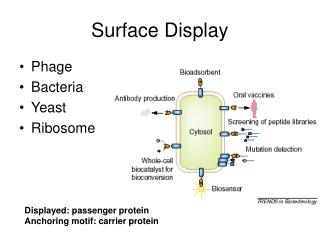
Surface Display. Phage Bacteria Yeast Ribosome . Displayed: passenger protein Anchoring motif: carrier protein. Phage Display. Panning.

Surface Display
E N D
Presentation Transcript
Surface Display • Phage • Bacteria • Yeast • Ribosome Displayed: passenger protein Anchoring motif: carrier protein
Phage Display Panning
Filamentous phages are flexible rods about 1 mm long and 6 nm in diameter, composed mainly (87% by mass) of a tube of helically arranged molecules of the 50-residue major coat protein pVIII22; there are 2700 copies in wild-type virions, encoded by a single phage gene VIII. Inside this tube lies the single-stranded viral DNA (ssDNA; 6407-8 nucleotides in wild-type strains). At one tip of the particle there are five copies each of the minor coat proteins pIII and pVI (genes III and VI, respectively); minor coat proteins pVII and pIX (genes VII and IX) are at the other tip.
Three rounds of panning againststreptavidin at constant stringency (0.5% Tween)
His-Pro-Gln (HPQ) as a consensus binding motif specific for thebiotin binding site of streptavidin
Epitope mapping of the anti-FLAG M2 monoclonal antibody. In order to determine which elements of the reported FLAG epitope sequence Asp-Tyr-Lys-Asp-Asp-Asp-Asp-Lys (DYKDDDDK) are required for antibody binding
Capture of the antibody by Protein A-agrose • Protein A is a cell wall protein deriving from Staphylococcus aureus which exhibits unique binding properties for IgG from a variety of mammalian species and for some IgM and IgA as well. It binds with the Fc region of immunoglobulins through interaction with the heavy chain.
Epitope mapping of an anti-β-endorphin monoclonal antibody with the Ph.D.-12 library
Cell surface display Gram (-) cell
Ice nucleation factor promoting ice forming
Ice Nucleation Protein (INP) OmpC
Gram(+) cell Staphylococall Protein A
Phage vs Ribosome Display Phage display:(purple) ribosome display: (green)
Ribosome display The DNA library constructs contain all the signals required for cell-free in vitro transcription and translation. The absence of a stop codon at the end of the coding sequence prevents the release of the mRNA and the nascent polypeptide from the ribosomes. Low temperatures and an elevated level of magnesium ions further stabilize the ternary mRNA–ribosome–polypeptide complex. An unstructured tether is added to the C-terminus of the constructs to enable correct folding of the nascent polypeptides outside the ribosome tunnel. After selection, mRNA molecules are recovered by reverse transcription (RT) and amplified by polymerase chain reaction (PCR).