Download
1 / 21
210 likes | 354 Vues
Basal Ganglia, Cerebellum and Movement. CBA 571 Structure of the Human Body (Chapter 10, Lundy-Ekman, emphasis on pp 190-215) Gib Willett, P.T., M.S., O.C.S., C.S.C.S. Associate Professor, UNMC P.T. Education. Objectives:.

E N D
Basal Ganglia, Cerebellum and Movement CBA 571 Structure of the Human Body (Chapter 10, Lundy-Ekman, emphasis on pp 190-215) Gib Willett, P.T., M.S., O.C.S., C.S.C.S. Associate Professor, UNMC P.T. Education
Objectives: • Explain the movement related functions of the basal ganglia and cerebellum. • Identify the basal ganglia and cerebellum in diagrams and on models. • Describe the neural pathways between these areas and the cerebrum which influence movement. • Describe most likely symptoms resultant from damage to a given region or pathway presented in this module.
Introduction • B.G. and cerebellum contain large collections of nuclei that modify movement on an ongoing basis • The motor cortex sends information to both areas and both respond back to cortex through the thalamus (gatekeeper to cortex)
Introduction • B.G. motor related signals to the motor cortex are inhibitory • Cerebellar motor related signals to the motor cortex are excitatory • Balance of these systems allows for smooth, coordinated movement
Basal Ganglia • What does ganglia usually refer to? • Collection of cell bodies outside the CNS • What are the Basal Ganglia (B.G.)? • B.G. are actually a collection of ganglia deep to the white matter of the cerebral cortex • Components of the B.G. include: • Caudate • Putamen • Globus pallidus • Subthalamic nucleus • Substantia nigria • There are more, but focus will be on the above five due to their involvement with movement Lentiform nucleus
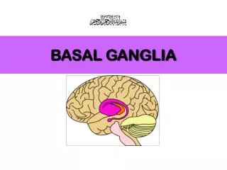
Basal Ganglia Location Where are the Globus Pallidus, Subthalamic Nucleus and Substantia Nigra? Thalamus B.G. component: Putamen Ventricles B.G. component: Caudate nucleus
Basal Ganglia: Location Frontal plane section of cerebrum
Basal Ganglia • Sequence movement • Regulate muscle tone and force • Two basal ganglia pathways influence movement • Promotion of certain movement patterns (synergies) • Inhibition of certain movement patterns (synergies) • Lesions of the basal ganglia result in disturbances of muscle tone and dyskinesias • Hyperkinesia • Hypokinesia
Basal Ganglia: Specifics • Caudate & Putamen receive majority of input from cortex (L-E only refers to the putamen) • Doorway into basal ganglia • Reciprocally interconnected with the substantia nigra
Basal Ganglia: Specifics • Substantia nigra • Pars compacta (SNpc)- receives input and sends information back • Produces dopamine which is critical for normal movement • See Parkinson’s Disease in L-E • Pars reticularis (SNpr)- receives input and sends it out to control head and eye movements
Basal Ganglia: Specifics • Globus pallidus • Most output from putamen goes to globus pallidus but not all…….(see figure 10-4, p223 in L-E for a more detailed diagram) • Both interna (not shown in L-E) and externa communicate with the subthalamic nucleus • Interna sends major inhibitory output from B.G. to cortex via thalamus • Interna also has output to midbrain to assist in postural control (see L-E diagram)
Cerebellum X-section showing cortex and deep nuclei Dorsal view • “Little brain” – outer cortex, inner white matter, deep nuclei • Coordinates movement: compares what you wanted to do (cortex), to what happens (proprioceptive feedback), and corrects the movement if needed. • Works ipsilaterally (cerebrum entirely contralateral)
Cerebellum Ventral view
Cerebellum • Summarized by 3’s • 3 highways leading in and out “peduncles” • Superior – connects to midbrain • Middle – connects to pons • Inferior – connects to medulla oblongata • 3 lobes – anterior, posterior and flocculonodular • 3 broad classes of human movements controlled for by the cerebellum (see L-E) • Equilibrium – vestibulocerebellum • Gross limb movements – spinocerebellum • Fine distal movements - cerebrocerebellum
Cerebellum • 3 input tracts • Spinocerebellar -proprioceptive feedback • Climbing fibers - feedback from ascending tracts via medulla • Pontine fibers - feedback from cerebral cortex • These fibers must cross then enter cerebellum
Cerebellum – “fun facts” • High density of neurons in cerebellar cortex results in cerebellum accounting for 1/10 of total brain volume but contains more than 50% of CNS neurons • Involved in motor learning process • Once cerebellum receives intent to move message, it provides information on movement direction, timing and force • Ballistic movements – too fast for feedback so predictions are made and modified via cerebellum based on experience (circuts change with repetition of an activity)
Cerebellar Lesions • Primarily coordination, proprioception and equilibrium related difficulties • Signs of cerebellar problems are manifested ipsilateral to the side of the lesion • Signs of cerebellar lesions include: • Hypotonia – flabby muscles, often pendulous reflexes • Dysmetria – past pointing, missing the mark • Dysdiadochokinesis – inability to make rapidly alternating movements
Cerebellar Lesions • Signs of cerebellar lesions continued: • Dysynergia - decomposition of movement ie. complex movements performed as a series of successive simple movements • Intention tremor – occurs with movement • Ataxia – stumbling gait • Alcohol abuse depresses cerebellar circuts (includes dysarthria) • Nystagmus – slow component towards the side of the lesion
Conclusion • Basal Ganglia – basically sequences movement and regulates muscle tone and force • Cerebellum – basically compares actual motor output to the intended movement and adjusts the motor output as necessary to meet movement expectations