Download
1 / 22
340 likes | 734 Vues
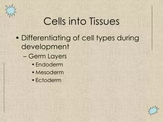
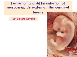
Directed differentiation of ES cells into cardiac mesoderm. Directed differentiation allows production of a desired cell type. Pluripotent. Multipotent. Differentiated cells. Heart muscle cells (or cardiomyocytes) derived by this process can be used for:

E N D
Directed differentiation allows production of a desired cell type Pluripotent Multipotent Differentiated cells Heart muscle cells (or cardiomyocytes) derived by this process can be used for: • 1. replacement of damaged tissue (e.g. following a heart attack). • 2. development of new therapies to treat chronic abnormalities of heart function (e.g. arrhythmia) by studying individual beating heart muscle cells in a dish. Ectodermal cell brain ES cell Mesodermal cell heart pancreas Endodermal cell 1
Diseases of heart muscle tissue Cardiac muscle – Tissue composed of individual cells, unlike skeletal myofibers with many nuclei – Cells coupled by gap junctions to enable rapid spread of electrical signals – Cells joined end-to-end by adhesive discs Diseases of cardiac muscles – Congestive heart failure, or ineffective pumping due to cardiomyocyte dysfunction, affects 4.8 million people in U.S. –Heart attack, also known as myocardial infarction –Heart arrhythmias, or abnormal activity (palpitations) – 1% of live births display a congenital heart defect
A four-chambered heart underliesdouble circulation (birds & mammals) • The circulatory system is: • Heart • Blood vessels • Blood cells • This system is the first functional unit that forms in the embryo. • Circulation provides nourishment to organs (nutrients and oxygen). • The heart is the main organ that pumps blood through the blood vessels. • The heart contains four chambers in birds and mammals (simpler in fish & amphibians). Right atrium Left atrium Atrioventricular valves Right ventricle Left ventricle 3
Heart muscle, to perform its unique function,is different from skeletal and smooth muscle 1. Cardiac muscle cells: • Have visible myofibrils • Occur only in heart • Involuntary movement • Cells joined by intercalating disks to allow rapid spread of electrical signals • 2. Skeletal muscle cells: • Elongated multinucleate cells called myofibrils. • Voluntary movements • 3. Smooth muscle cells: • Individual, spindle-shaped mononuclear cells • Found in gut, blood vessels and ducts of glands • Involuntary movements Three types of muscle cells 4
How are cardiomyocytes generated during development? Lineage restrictions Pluripotent Multipotent Differentiation Ectodermal cell ? ? ES cell Mesodermal cell Multipotent cardiovascular progenitor Cardiomyocyte Endodermal cell 5
Heart, blood and blood vessels are derived from the lateral mesoderm Heart, circulatory system, smooth muscle 6 Skeletal muscles
Secreted factors specifyheart-forming mesoderm in vivo Lineage restrictions Differentiation FGF8 Cardiogenic mesodermal cell Anterior lateral plate mesodermal cell Cardiomyocyte Cerberus BMPs Wnts Lateral plate mesodermal cell Noggin Posterior lateral plate mesodermal cell Hemangiogenic mesodermal cell Blood cells 9
Distinct cell signaling pathways induce cardiomyocyte-specific genes Cell signaling pathway Gene expression Heart muscle cell (Cardiomyocyte) BMPs BMP-RII Progenitor cell OFF OFF ON Heart muscle cell (Cardiomyocyte) Myosin light chain 2 gene 10
Markers of the cardiomyocyte progenitor lineage Cardiomyocyte lineage Differentiation Bry Nkx2.5+ / Isl-1+ Nkx2.5+ / c-Kit+ 1st heart field progenitor Left ventricle Cardiogenic mesodermal progenitor Mesodermal Precursor Nkx2.5+ / Isl-1+/ GATA4+ Cardiac progenitor Remainder of heart Nkx2.5+ / Isl-1+/ GATA4+/ Flk-1+ Isl-1+/ Flk-1+ Multipotent cardiovascular progenitor (2nd heart field) Blood vessels and blood cells Vascular progenitor 11
Sources of cells for cardiomyocyte replacement therapies: the dish Directed differentiation Transplantation & engraftment Stable formation of mural grafts
Sources of cells for cardiomyocyte replacement therapies: the heart
Directed differentiation protocol for mouse ES cells into cardiomyocytes - LIF + serum - LIF + serum - LIF + serum Day 5-7 Nkx2.5+ progenitors Day 7-10 beating cardiomyocytes Day 2 EBs mES cells Wnt/BMP/Activin and serum are often used to induce cardiomyocyte precursors 2 - 4 days 4 days 2 days ES cells EBs Nkx2.5+ cardiomyogenic progenitors Beating cardiomyocytes 15
Directed differentiation of mouse ES cells produces vascular and cardiac lineages Isl-1+/ Flk-1+ Nkx2.5+ / Isl-1+/ Gata4+ Vascular progenitors Cardiac progenitors Fraction of Flk-1+ progenitors at day 3-5 Fraction of Nkx2.5+ progenitors at day 5 % % % 3.84 % 16 Nkx2.5::eGFP
Isolated heart muscle cellsbeat spontaneously • Directed differentiation of ES cells creates specialized beating heart cells in vitro. • How do heart muscle cells (or cardiomyocytes) beat in a dish? • 1. Express cardiac troponin T (cTnT). • 2. Some cells become node cells (HCN4+) that beat spontaneously. • 3. These cells express Ca++ ion channels that confer contractile properties. 17
Directed differentiation protocol for human ES cells to cardiomyocytes - bFGF + 20% FBS - bFGF + 20% FBS - bFGF + 20% FBS BMP-4 Day 7 Nkx2.5+ progenitors Day 11 beating cardiomyocytes Day 4 hEBs hES cells Wnt/BMP/Activin and serum are often used to induce cardiomyocyte precursors 4 days 3 days 4 days 20% FBS BMP-4 12.5-25 ng/ml 20% FBS 20% FBS hES cells Nkx2.5+ cardiomyogenic progenitors Human embryoid bodies Beating cardiomyocytes 18
Rodent model of heart attack to test stem cell-based repair of heart tissue • Sources for stem/progenitor cells that might be used to repair tissue following a heart attack: • ES cells • Cardiac stem cells • Muscle stem cells • Mesenchymal cells from adult bone marrow • Endothelial progenitors • Umbilical cord blood cells • Rats, mice and pigs are commonly used to test whether cells can repair hearts • Conditions mimicking those of a heart attack are achieved in animal models by ligation of the coronary artery
Stem cells directly or indirectlyrepair damaged heart tissue 20
Summary Directed differentiation of ES cells into heart muscle cells is the production of beating cardiomyocytes in a dish using defined factors. The factors used are crucial for generating these muscle cells during normal embryonic development. Formation of the mammalian heart involves complex tissue rearrangements as well as precise cell differentiation. Similar experimental conditions lead to directed differentiation of cardiomyocytes from either mouse or human ES cells. A wide variety of sources may provide cells that are effective for repairing heart muscle tissue after is has been damaged due to a heart attack. 21