Download
1 / 36
360 likes | 402 Vues
This lecture covers the fundamentals of anatomical terminology, skeletal classification, body planes, cavities, regions, and terms of position and movement. Understanding these concepts is crucial in the study of human anatomy.

E N D
Anatomical Terminology&Skeletal System DR JAMILA EL MEDANY
OBJECTIVES At the end of the lecture, students should be able to: • Define the word “Anatomy” • Enumerate the different anatomical fields • Describe the anatomical position • Describe different anatomical terms of position & movements as well different anatomical planes • Classify bones according to shape, structure & development • Enumerate bones of axial & appendicular skeleton
ANATOMY (to Cut) • The science which deals with the study of the structure and shape of the body & body parts, and their relationships to one another • It is divided into: • Gross Anatomy: Study of human body with naked eye • Microscopic Anatomy (Histology): Study of fine structures (cells & tissues) of the human body with the help of microscope • Developmental Anatomy ( Embryology) • Radiological Anatomy • Cross-sectional Anatomy • Applied Anatomy • Surgical Anatomy
The Language of Anatomy(Anatomical Terminology) • To prevent misunderstanding, a special set of terms are used to describe the identification and location of body structures • To accurately describe body parts, the body is in a standard position called the Anatomical Position, in which: • Body is erect • Arms hanging by the side • Palms facing forward • Feet are parallel
PLANES OF THE BODY To do a Section (cut) through the body wall or an organ, it is made along • an Imaginary Line (PLANE). • The body has Three Imaginary Planes (sections ) • that lie at right angles to one another (in the anatomical position). • 1.Median sagittal. • 2. Coronal. • 3.Horizontal (Transverse).
MEDIAN (MidSagittal )PLANE • It is a Vertical plane. • It passes through the Center (Midline)of the body. • It divides the body into Right andLefthalves.
CORONAL (FRONTAL) PLANE • It is a Vertical plane. • It divides the body into : • Anteriorand Posteriorparts.
HORIZONTAL (TRANSVERSE ) PLANE • It is also called Cross Section. • It divides the body into : • Upperand Lowerparts.
Terms of Regions • Cranial (Cephalic) • Cervical • Thoracic • Abdominal • Pelvic • Planter • Palmer
Body Cavities The body has two sets of internal cavities that lodge and protect the organs. These are Dorsal & Ventral. • Dorsal body cavityhas two subdivisions, which are continuous with each other: • Cranial cavity: space inside the bony skull, contains brain • Spinal cavity: space inside the vertebral column, contains spinal cord • Ventral body cavity has two subdivisions, which are separated from each other by the diaphragm. • Thoracic cavity: lies superior to diaphragm, contains heart and lungs • Abdominopelvic cavity: lies below the diaphragm, contains stomach, intestine, urinary bladder, liver, reproductive organs, rectum, etc.
Abdominopelvic regions The Abdominopelvic area is divided into 9 regions by 2 vertical & 2 horizontal lines or planes Objective: To locate the different organs in each region
TERMS OF POSITION • Anterior: Front of the body. • Posterior: Back of the body. • (HAND) : • Anterior:Palmar. Posterior: Dorsal . • (FOOT) : • Anterior:Planter . • Posterior:Dorsal. • Medial :Nearer to the median plane of the body. • Lateral : Away from the median plane.
TERMS OF POSITION • Superior (Above): Toward the head end (upper) part of the body. • Inferior(Caudal) :Toward the lower part ofthe body. • Supine : • The body lies on the back. • Prone : • The face is downwards.
Proximal :Close to the point of attachment of a limb to the body trunk. • Distal :Farther from the the point of attachment of a limb to the body trunk. • Superficial (External) : Toward or at the body surface. • Deep (Internal):Away from the body surface or the center of a cavity. • .
TERMS OF MOVEMENT. • A. Flexion: • Usually an Anterior movement (Except. • in the knee joint). • It Decreases the angle of the joint (brings two • bones closer together). • B. Extension: • Usually a Posteriormovement. • Straightening of the joint. • It Increases the angle or distance between two bones.
Movements In the Coronal (frontal) plane: • 1. Abduction: • Movement of a limb Away from the midline of the body • 2. Adduction: • Movement of a limb Towardthe midline of the body. 3. Lateral flexion: Side Movement of the trunk
Circumduction • It is Combination of: • Flexion. • Extension. • Abduction. • Adduction
ROTATION • Medial: • The anterior surface of the part faces medially. • Lateral : • The anterior surface of the part faces laterally.
Opposition: bringing tips of fingers and thumb together as in picking something up Supination: • Lateral rotation of the forearm. • The palm faces Anteriorly. • The radius and ulna are Parallel. • Pronation: • Medial rotation of the forearm. • The palm faces Posteriorly • The radius Crossesthe ulna and the two bones form an X.
Planter Flexion: • Depressing the foot(down ). • Movement with pointing the toes. • Dorsiflexion • Up movement of the foot • (Standing on the heels) • Inversion : • The sole faces in a Medialdirection. • Eversion : • The sole faces in a Lateral direction.
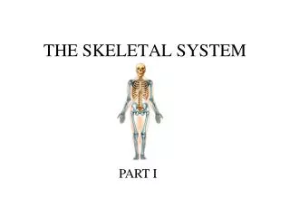
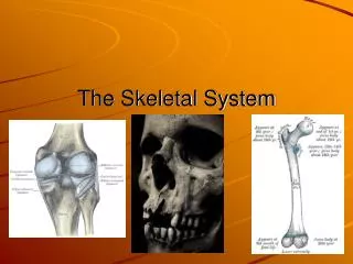
Skeletal System Includes: • Bones • Joints (articulations)
Functions of Bones Support of the body organs Protection of soft body organs Attachment of muscles Movementof the body as a whole, or of the body parts Storage of fat and minerals e.g. calcium and phosphorus Blood cell formation
Classification of Bones Bones are classified on the bases of their: • 1. Shape: • Long, Short, Flat, Irregular • 2. Structure: Compact & Spongy • 3. Development: Membranous & Cartilagenous
Gross Structure of a Long Bone • Each long bone has: • A long cylindrical shaft called the ‘diaphysis’. • Two ends called the ‘epiphyses’ • The region at the junction of diaphysis and epiphysis is called ‘metaphysis’
Diaphysis (Shaft) • Composed of compact bone • Covered on its external surface by a fibrous connective tissue membrane called the periosteum. • Has a cavity called the marrow cavity. In adults, the marrow cavity is a storage area for fat and contains yellow marrow. In infants, it containsred marrowand is the site of blood cells formation
Epiphyses Each epiphysis is composed of spongy bone, lined by a thin layer of compact bone. Its external surface is covered by a layer of hyaline cartilage called the articular cartilage Articular cartilage provides smooth slippery surface that decreases friction at joint surfaces Metaphysis • It contains a thin plate of cartilage called the epipyseal plate, that is responsible for the lengthwise growth of the long bones.
Role of Periosteum • Protects the bone • Gives attachment to muscles • Carries blood vessels and nerves to bone • Deposits new bone on the surface thus increases the girth of bone Growth of bone • Increase in length: epiphyseal plates • Increase in girth: periosteum
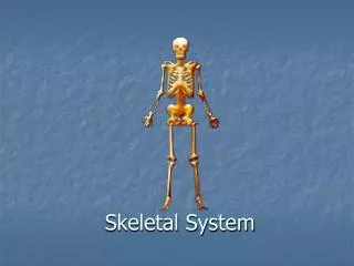
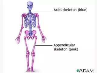
The Skeleton • There are 206 bones in our body, arranged to form the body framework called, the skeleton • The skeleton is perfectly adapted to the functions ofbody protectionandmotion • It is subdivided into two divisions: • The Axial skeleton, the bones that form the longitudinal axis of the body • TheAppendicular skeleton, the bones of limbs and girdles
The Axial Skeleton consists of the: Skull bones Vertebral column Sternum Ribs • The Appendicular Skeletonconsists of the bones of the : • Pectoral & Pelvic Girdles, connect the bones of the limbs to the axial skeleton • Upper Limb • Lower Limb
Skull bones • Formed of two sets of bones: • Cranium: • Encloses and protects the brain. • Consists of the following bones: • Frontal • Parietal • Temporal • Sphenoid • Occipital • Facial bones: • Form the skeleton of the face • Consists of the following bones: • Maxilla • Mandible • Zygomatic • Nasal
Vertebral column • Forms the axial support of the body • Is a flexible curved structure, formed of 33 irregular bones, the (vertebrae) • Running through its cavity is the spinal cord • Is divided into 5 regions: • Cervical: 7 vertebrae • Thoracic: 12 vertebrae • Lumbar: 5 vertebrae • Sacral: 5 vertebrae fused to from a triangular bone called sacrum • Coccygeal: 4 vertebrae fused to form a small bone called coccyx
Sternum • Flat bone • Has three parts: manubrium, body and xiphoid process Ribs • Number: 12 pairs • All ribs articulate with vertebrae • Only upper 7 pairs articulate with sternum
Bones of the Girdles • Pectoral Girdle: Bones connecting the upper limb with the axial skeleton • Clavicle • Scapula • Pelvic Girdle: Bones connecting the lower limb with the axial skeleton • Two hip bones
Bones of the Upper Limb • Bone of arm: humerus • Bones of forearm: radius (lateral) & ulna (medial) • Bones of hand: • 8 carpal bones • 5 metacarpal bones • 14 phalanges: 2 for thumb & 3 for each of medial 4 fingers
Bones of the Lower Limb • Bone of thigh: femur • Bones of leg: fibula (lateral) & tibia (medial) • Patella • Bones of foot: • 8 tarsal bones • 5 metatarsal bones • 14 phalanges: 2 for big toe & 3 for each of lateral 4 toes