Download
1 / 76
760 likes | 814 Vues
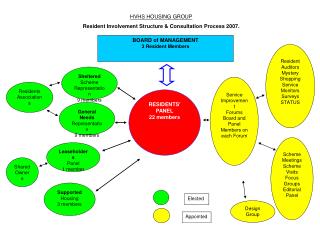
This article explores the causes and management of dysphagia, encompassing grades, common causes, and clinical presentations. With a focus on dysphagia due to various disorders and conditions affecting the esophagus, it delves into the anatomy and physiology of swallowing to aid in comprehensive understanding.

E N D
Dysphagia – Causes& ManagementDr.Nayana.V.G MBBS,MS Senior ResidentDept of ENT
Esophagus • BRIEF ANATOMY : • Esophagus is a tubular structure • about 10 inches • continuous above with laryngeopharynx opposite 6th cervical vertebra • passes through diaphragm at the level of 10th thoracic vertebra where it joins the stomach in the abdomen
upper 3rd Inferior thyroid artery Inferior thyroid vein • BLOOD SUPPLY OF ESOPHAGUS: middle 3rd Descending thoracic aorta azygos vein lower 3rd left gastric vein Left gastric artery
AREAS OF NARROWING IN ESOPHAGUS: • The aortic arch • The left main bronchus • The diaphragm
Physiology of swallowing Oral Phase. Pharyngeal phase Closure of nasopharynx Closure of oral cavity Closure of larynx Pharyngeal contractions Esophageal Phase In resting both UES and LES are contracted to prevent reflux .
GRADES OF DYSPHAGIA: mild dysphagia: only solid things can't be swallowed moderate dysphagia: solid & semi solid thing can't be swallowed severe dysphagia: even liquids can't pass DYSPHAGIA • DEFINITION OF DYSPHAGIA: Medical term means any difficulty in swallowing regardless of the cause • GRADES OF DYSPHAGIA: • mild dysphagia: only solid things can't be swallowed • moderate dysphagia: solid & semi solid thing can't be swallowed • severe dysphagia: even liquids can't pass
COMMON CAUSES OF DYSPHAGIA: • CONGENITAL • ACQUIRED
1.CONGENITAL: esophageal atresia
Pre esophageal etiologies ORAL PHASE • Disturbances in mastication • Disturbances in Lubrication • Tongue disorders • Palatal Defects • Lesion of buccal cavity
Pharyngeal Causes a)Obstructive lesion of pharynx b) Inflammatory condition c)Paralytic condition d)Spasmodic condition
ESOPHAGEAL ^ In the lumen Foreign body Food bolus strictues ^ In the wall Hyomobility disordes- Achalasia, Scleroderma Chagas’ disease Hyermobility Disorders Tumour of the esophagus GERD Plummer Vinson syndrome Pharyngeal pouch ^ Outside the wall Pressure of the enlarged lymph nodes Thoracic aortic aneurysm Bronchial carcinoma Retrosternal goitre ^ Neuromuscular disorders Myesthenia gravis Bulbar palsy Bulbar poiliomylitis
Associated symptoms: • Difficulty in chewing or bolus preparation • Excessive drooling • Chocking or regurgitation • Food sticking • Symptoms aggravated by : • Aspiration pneumonia • Weight loss • Compromised nutritional status • Or any combination of these
Benign stricture Etiology – Trauma,Burns,Ulceration,surgery C/F – Dysphagia Invstigation – Ba.Swallow, OGD scopy Rx : BOUGIE Dilatation, Gastrostomy, surgery
Hiatus Hernia • 2 Types Sliding – reflux symptoms Paraesophageal – DOE Ba.swallow Rx:Surgery
Plummer –Vinson syndrome “Patterson Brown kelly Syndrome” Sideropenic Dysphagia C/F : Anemia, Glossitis, Angular stomatits,Koilonychia,Achlorhydria, Splenomegaly,esophageal web
Females , >40 yrs • 10% a/w Sqcc of Post cricoid area • Dx- Ba.Swallow and UGI scopy • Rx: parenteral iron • Esophageal Bougies
Motility Disorders • Hypermotility Disorders • Cricopharyngeal Spasm: Failure of UES to relaxation B) Diffuse esophageal spasm- Non peristaltic contraction of body of esophagus with normal sphincteric contractions Corkscrew esophagus Manometry – NAD Rx: Dilatation : Surgery ( MYOTOMY from arch of aorta to LES)
Corkscrew esophagus • Manometry – NAD • Rx: Dilatation : Surgery ( MYOTOMY from arch of aorta to LES)
C) Nut Cracker Esophagus Strong high amplitude peristaltic esophageal contractions C/F : Dysphagia and substernal pain Diagnosis: Manometry; Ba.swallow – NAD Rx: Medical Treatment (CCB, Nitrates) Botulinum Toxin Surgery
Achalasiacardia Definition Primary esophageal motility disorder characterized by: 1) Absence of peristalsis, 2) Elevated pressure of the LES, 3) Failure of the LES to relax during swallowing, Causing functional obstruction at the gastroesophageal junction.
What is the most likely diagnosis? Achalasia of the Cardia WWW.MD4A.NET
Clinical picture of Achalasia History • Dysphagia (most common) • Regurgitation • Chest pain • Heartburn • Weight loss
Details • 25-50% report episodes of retrosternal chest pain. • 80-90% experience spontaneous regurgitation. • some patients may present with signs or symptoms of pneumonia or pneumonitis. • Patients with achalasia are at increased risk for esophageal cancer Physical: Physical examination is noncontributory.
INVESTIGATIONS Lab Studies • Laboratory studies are noncontributory. Imaging Studies • Ba.Swallow: Bird’s Beak. • EGD: Normal or dilated esophagus. • Manometry The radiologic examination of choice in the diagnosis of achalasia is a barium swallow study performed under fluoroscopic guidance.
Normal Barium Swallow Bird’s Beak Birds bak xray
Diagnosis of achalasia supported by the results of radiologic studies must always be confirmed by 1) Performing (EGD) to rule out cancer of the gastroesophageal junction or fundus, and 2) Esophageal manometry.
MANAGEMENT • The goal of therapy for achalasia is to relieve symptoms by eliminating the outflow resistance caused by the hypertensive and nonrelaxing LES. • Medical Management • Surgical Management
Medical Management • Botulinum toxin: intrasphincteric injection Only 30% of patient's treated endoscopically still have relief of dysphagia 1 year after treatment. (50% re-treat in 9 months vs 10% balloon)
b) Pharmacologic therapy: relax the smooth muscle of the LES. • Calcium channel blockers - Nifedipine and verapamil • Anticholinergic agents - Cimetropium bromide • Nitrates - Isosorbidedinitrate • Opioids - Loperamide
Surgical Management • Pneumatic dilatation: Dilation of LES with a bougie ( flexible cylindrical instrument ) Most commonly performed by using pneumatic balloons. The success rate is 70-80%, and the perforation rate is approximately 5%.
Approximately one half of patients experience recurrent symptoms within 5 years. Complications Failures Perforation Gastro esophageal reflux
b)Esophageal (Heller) myotomy: ^ Heller myotomy – cardiomyotomy and dividing the muscle of the lower end of the esophagus and the upper stomach down the mucosa. ^ Heller myotomy is the appropriate treatment for patients in whom pneumatic dilatation fails. ^ The laparoscopic approach appears to be most appropriate.
Under general anesthesia, make a controlled division of the muscle fibers (myotomy) of the lower esophagus (5 cm) and proximal stomach (1.5 cm) followed by a partial fundoplication to prevent reflux . • Patients in whom surgery fails may need a dilatation, a second operation, or removal of the esophagus (ie, esophagectomy).
Esophageal Tumor • Benigh Tumors: * LIOMYOMA * POLYP • Malignant Tumors
Benign Tumors • RARE • The commonest is LIOMYOMA. • Dysphagia , bleeding. • Remove it .
Esophageal Carcinoma • Oesophageal carcinoma is becoming more common, unlike the trend observed in gastric carcinoma. • Squamous carcinoma typically arises in the upper 2/3 of the oesophagus • Adenocarcinoma usually arises in a region of specialised columnar epithelium (SCE) metaplasia in the lower 1/3 of the oesophagus - Barrett's oesophagus.
Clinical Features • Dysphagia is the most frequently presented feature of oesophageal carcinoma : • Patient may recount a short history of progressive dysphagia, initially affecting solids only, but gradually affecting the swallowing of fluids.
Clinical Features • The level at which difficulty in swallowing is encountered may be identifiable by the patient. • Short history of dysphagia in an elderly male is almost certainly carcinoma of the oesophagus or the cardia of the stomach.
Clinical Features • Other local features may include : • Regurgitation of food or blood-stained vomit • Aspiration pneumonia • Pain : • classically, retrosternally and in the interscapular region • it may radiate to the jaws and arms
Clinical Features • General manifestations of malignant disease may include : • Loss of Weight • Anorexia • Anemia • Coughing • Secondary deposits occur in: • lymph nodes • liver causing jaundice