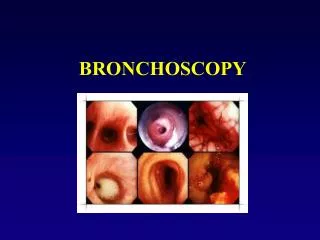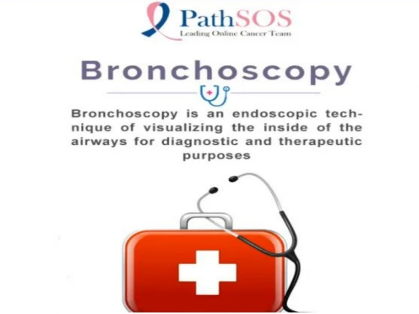
BRONCHOSCOPY
This article explores bronchoscopy as a vital clinical tool for direct visualization of the airways. It covers both rigid and flexible bronchoscopy, detailing various instruments, techniques, and indications for procedures. From foreign body removal to biopsy and therapeutic interventions, the paper highlights the evolution of bronchoscopic practices from the 1980s to present. Key information on anesthesia, anatomical surveys, and potential complications is provided, making this an essential resource for healthcare professionals in pulmonary medicine and surgery.

BRONCHOSCOPY
E N D
Presentation Transcript
IN THE NAME OF GOD BRONCHOSCOPY Mehdi hadadzadeh, MD Cardiovascular surgeon University of yazd
BACKGROUND • Allows direct visualization of the airways • Rigid and flexible instruments • Clinical tool • Airway anatomy • Airway sampling • Therapeutic • Research tool
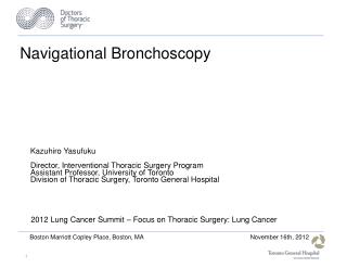
ORIGINS • Until the 1980’s, only rigid instruments were widely used • Multiple generations of adult and pediatric flexible bronchoscopes now • Widely used in adult and pediatric pulmonary medicine now
RIGID BRONCHOSCOPY • Generally performed by ENT’s and surgeons • Procedure oriented • Foreign body removal • Biopsies • Granuloma/polyp removal • Laser • Stent placement • Visualization for future surgery
INSTRUMENTS • Rigid bronchoscopes • Hollow metal tube • Glass rod telescope
FLEXIBLE BRONCHOCSOPY • Examination of the entire respiratory anatomy, nose to bronchi • Able to pass through an endotracheal tube or tracheostomy tube
INSTRUMENTS • Flexible instruments • Fiberoptic bronchoscopes • 2.2mm ultrathin • 2.8mm/1.2mm suction channel • 3.4mm/1.2mm suction channel • 4.4mm/2.0mm suction channel • 4.9mm/2.2mm suction channel • 5.9mm/3.0mm suction channel
Fiberoptic bronchoscope 2.8mm diameter
Pediatric videoscope 3.8mm diameter
INDICATIONS • Atelectasis • Recurrent pneumonia • Chronic cough • Persistent/unexplained wheeze • Hemoptysis • Suspected airway compression/obstruction • Stridor • Upper airway obstruction • Suspected aspiration • Evaluation of tracheostomies
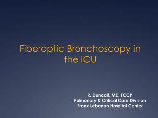
TECHNIQUE • Anesthesia • Best accomplished in the operating room • May be performed bedside in an ICU setting • Continuous monitoring • Light anesthesia--allows continued spontaneous breathing • May be done with conscious sedation in older individuals
TECHNIQUE • Insertion • Nasal • Endotracheal tube • Tracheostomy tube • Appropriate topical anesthesia and lubrication
TECHNIQUE • Anatomical survey • Nasal passages • Pharynx • Larynx • Trachea • Bronchi • Examine all before any other procedures
TECHNIQUE • Additional procedures • Bronchoalveolar lavage • Brushings • Bronchial biopsy • Transbronchial biopsy • Laser • Others: cryotherapy, stent placement, foreign body removal, needle biopsy
BRONCHOALVEOLAR LAVAGE • Small aliquots of sterile normal saline instilled into the airway • Removed by suctioning • Samples distal bronchial and alveolar surfaces
Thoracoscopy insertion of an endoscope, a narrow-diameter tube with a viewing mirror or camera attachment, through a very small incision (cut) in the chest wall
Thoracoscopy two or three small incisions in the chest wall, often between the ribs examine the pleura, lungs, and mediastinum to obtain tissue for testing purposes general anesthesia
Indications assess lung cancer take a biopsy for study determine the cause of fluid in the chest cavity introduce medications or other treatments directly into the lungs treat accumulated fluid, pus (empyema), or blood in the space around the lungs
The risks of thoracoscopy • Wound infection • Bleeding • Air leak through the lung wall, requiring a longer hospital stay • Pain or numbness at the incision site • Inflammation of the lungs (pneumonia)
thoracoscopy v;s thoracotomy avoids many of the complications of open chest surgery reduces pain, hospital stay recovery time.
Preparation • chest X-ray • electrocardiogram (if you are over age 35) • various blood tests • arterial blood gas • pulmonary function test • fast for 12 hours before the procedure. • General anesthesia • preparations for chest surgery
Transthoracic Needle Biopsy to evaluate peripheral lung nodules or masses; hilar, mediastinal, and pleural abnormalities; and undiagnosed infiltrates or pneumonias when bronchoscopy is contraindicated or nondiagnostic
diagnosis of cancer with > 95% accuracy. Needle biopsy yields an accurate diagnosis in benign processes only 50 to 60% of the time.
Complications: • hemoptysis (10 to 25%) • pneumothorax (10 to 37%) • parenchymal hemorrhage • air embolism • subcutaneous emphysema.
Mediastinoscopy surgical procedure to examine the inside of the upper chest between and in front of the lungs (mediastinum).
replaced by other biopsy methods that use computed tomography (CT), echocardiography, or bronchoscopy to guide a biopsy needle to the abnormal tissue
Indications • Detect problems of the lungs and mediastinum, such as sarcoidosis. • Diagnose lung cancer or lymphoma (including Hodgkin's disease). Mediastinoscopy is often done to check lymph nodes in the mediastinum before considering lung removal surgery to treat lung cancer. Mediastinoscopy can also help your doctor recommend the best treatment (surgery, radiation, chemotherapy) for lung cancer. • Diagnose certain types of infection, especially those that can affect the lungs (such as tuberculosis).
limitations • previous mediastinoscopy or open-heart surgery • A history of neck problems or a neck injury • Any physical problems of chest(congenital). • Recently radiation therapy to the neck or chest.
risks • puncturing the esophagus, trachea, or blood vessels . • In some circumstances, this can lead to potentially fatal bleeding.