VIRTUAL BRONCHOSCOPY-CLINICAL CASES
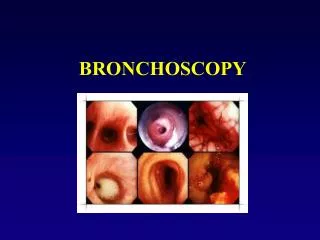
VIRTUAL BRONCHOSCOPY-CLINICAL CASES. Normal VB Views n. 1st 4 images show normal carina, right upper and middle lobe bronchus, R lower lobe bronchus and L lower lobe segmental bronchi respectively. Sq Ca with occluded LLL bronchus.

VIRTUAL BRONCHOSCOPY-CLINICAL CASES
E N D
Presentation Transcript
Normal VB Viewsn 1st 4 images show normal carina, right upper and middle lobe bronchus, R lower lobe bronchus and L lower lobe segmental bronchi respectively.
Sq Ca with occluded LLL bronchus Very poorly tolerated FOB. Left side not well visualised but washings proved Sq Ca ( mass seen on 1st CT image). VB showed occluded LLL bronchus ( seen on next 2 images). Patient received subsequent radiotherapy because of severe breathlessness.
Known Sq Ca • Massive haemoptysis. Too ill for FOB. VB shows markedly narrowed LMB ( seen also on multi-planar reconstuction [MPR] image). Radiotherapy planned. This patient later shown to also have a TOF.