Neural Tissue: Glial Cells and Neurons Overview
490 likes | 525 Vues
Explore the intricate world of neural tissue, from glial cells like astrocytes and oligodendrocytes to the functions and structures of neurons. Discover the role of neural tissue in coordinating bodily activities.

Neural Tissue: Glial Cells and Neurons Overview
E N D
Presentation Transcript
Two organ systems coordinate and direct activities of body • Nervous system • Swift, brief responses to stimuli • Endocrine system • Adjusts metabolic operations • Directs long-term changes
Anatomical Classification of the Nervous System • Central Nervous System • Brain and spinal cord • Peripheral Nervous System • All neural tissue outside CNS
Functional divisions of nervous system • Afferent • Sensory information from receptors to CNS • Efferent • Motor commands to muscles and glands • Somatic division • Voluntary control over skeletal muscle • Autonomic division • Involuntary regulation of smooth and cardiac muscle, glands
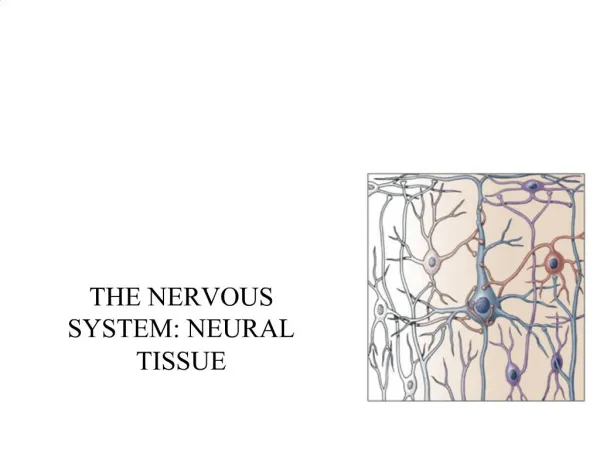
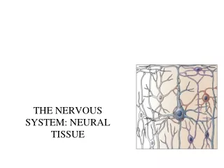
Cells in Nervous Tissue • Neurons • Neuroglia
Neuroglia (Glia) • about half the volume of cells in the CNS • smaller than neurons • 5 to 50 times more numerous • do NOT generate electrical impulses • divide by mitosis • two types in the PNS • Schwann cells • Satellite cells • Four types in the CNS • Astrocytes • Oligodendrocytes • Microglia • Ependymal cells
Astrocytes • Largest of glial cells • Star shaped with many processes projecting from the cell body • Help form and maintain blood-brain barrier • Provide structural support for neurons • Maintain the appropriate chemical environment for generation of nerve impulses/action potentials • Regulate nutrient concentrations for neuron survival • Regulate ion concentrations - generation of action potentials by neurons • Take up excess neurotransmitters • Assist in neuronal migration during brain development • Perform repairs to stabilize tissue
Oligodendrocytes • Most common glial cell type • Each forms myelin sheath around the axons of neurons in CNS • Analogous to Schwann cells of PNS • Form a supportive network around CNS neurons • fewer processes than astrocytes • round or oval cell body
Microglia • few processes • derived from mesodermal cells that also give rise to monocytes and macrophages • Small cells found near blood vessels • Phagocytic role - clear away dead cells • protect CNS from disease through phagocytosis of microbes • migrate to areas of injury where they clear away debris of injured cells - may also kill healthy cells
Ependymal Cells • epithelial cells arranged in a single layer • range in shape from cuboidal to columnar • Form epithelial membrane lining cerebral cavities (ventricles) & central canal - that contain CSF • Produce & circulate the cerebrospinal fluid (CSF) found in these chambers • CSF = colourless liquid that protects the brain and SC against chemical & physical injuries, carries oxygen, glucose and other necessary chemicals from the blood to neurons and neuroglia
PNS: Satellite Cells • Flat cells surrounding PNS cell bodies • Support neurons in the PNS
PNS: Schwann Cells Neurilemma • each cell surrounds multiple unmyelinated PNS axons with a single layer of its plasma membrane • Each cell produces part of the myelin sheath surrounding an axon in the PNS • contributes regeneration of PNS axons
Representative Neuron -neurofilaments or neurofibrils give cell shape and support - bundles of intermediate filaments -microtubules move material inside cell -lipofuscin pigment clumps (harmless aging) - yellowish brown 1. cell body or soma -single nucleus with prominent nucleolus -Nissl bodies -rough ER & free ribosomes for protein synthesis -proteins then replace neuronal cellular components for growth and repair of damaged axons in the PNS
Neurons 2. Cell processes = dendrites (little trees) - the receiving or input portion of the neuron -short, tapering and highly branched -surfaces specialized for contact with other neurons -cytoplasm contains Nissl bodies & mitochondria
3. Cell processes = axons • Conduct impulses away from cell body-propagates nerve impulses to another neuron • Long, thin cylindrical process of cell • contains mitochondria, microtubules & neurofibrils - NO ER/NO protein synth. • joins the soma at a cone-shaped elevation = axon hillock • first part of the axon = initial segment • most impulses arise at the junction of the axon hillock and initial segment = trigger zone • cytoplasm = axoplasm • plasma membrane = axolemma • Side branches = collaterals arise from the axon • axon and collaterals end in fine processes called axon terminals • Swollen tips called synaptic end bulbs contain vesicles filled with neurotransmitters
Structural Classification of Neurons • Based on number of processes found on cell body • multipolar = several dendrites & one axon • most common cell type in the brain and SC • bipolar neurons = one main dendrite & one axon • found in retina, inner ear & olfactory • unipolar neurons = one process only, sensory only (touch, stretch) • develops from a bipolar neuron in the embryo - axon and dendrite fuse and then branch into 2 branches near the soma - both have the structure of axons (propagate APs) - the axon that projects toward the periphery = dendrites
Structural Classification of Neurons • Named for histologist that first described them or their appearance • Purkinje = cerebellum • Renshaw = spinal cord • others are named for shapes e.g. pyramidal cells
Functional Classification of Neurons • Sensory (afferent) neurons • transport sensory information from skin, muscles, joints, sense organs & viscera to CNS • Motor (efferent) neurons • send motor nerve impulses to muscles & glands • Interneurons (association) neurons • connect sensory to motor neurons • 90% of neurons in the body
Terms to know • membrane potential = electrical voltage difference measured across the membrane of a cell • resting membrane potential = membrane potential of a neuron measured when it is unstimulated • results from the build-up of negative ions in the cytosol along the inside of the neuron’s PM • the outside of the PM becomes more positive • this difference in charge can be measured as potential energy – measured in millivolts • polarization • depolarization • repolarization • hyperpolarization
The electric potential across an axonal membrane can be measured • the differences in positive and negative charges in and out of the neuron can be measured by electrodes = resting membrane potential -ranges from -40 to -90 mV
ion channels in the PM of neurons and muscles contributes to their excitability • when open - ions move down their concentration gradients • channels possess gates to open and close them • two types: gated and non-gated Ion Channels 1. Leakage (non-gated) or Resting channels: are always open, contribute to the resting potential -nerve cells have more K+ than Na+ leakage channels -as a result, membrane permeability to K+ is higher -K+ leaks out of cell - inside becomes more negative -K+ is then pumped back in 2. Gated channels: open and close in response to a stimulus A. voltage-gated: open in response to change in voltage - participate in the AP B. ligand-gated: open & close in response to particular chemical stimuli (hormone, neurotransmitter, ion) C. mechanically-gated: open with mechanical stimulation
Action Potential • Resting membrane potential is -70mV • triggered when the membrane potential reaches a threshold usually -55 MV • if the graded potential change exceeds that of threshold – Action Potential • Depolarization is the change from -70mV to +30 mV • Repolarization is the reversal from +30 mV back to -70 mV) • action potential = nerve impulse • takes place in two stages: depolarizing phase (more positive) and repolarizing phase (more negative - back toward resting potential) • followed by a hyperpolarizing phase or refractory period in which no new AP can be generated http://www.blackwellpublishing.com/matthews/channel.html
The action potential 9. • 1. neuron is at resting membrane potential (resting MP) • 2. neuron receives a signal • Neurotransmitter (NT) • 3. NT binds ligand-gated sodium channel • 4. LGNa channel opens • 5. Na flows into neuron = depolarization • Inside of neuron (i.e. MP) becomes more positive • 6. if neuron depolarizes enough to Threshold = Action Potential (AP) • 7. 1st stage of AP – opening of voltage-gated Na channels • 8. even more Na flows in through VGNa channels = BIG depolarization • Membrane potential goes from negative to positive • 9. closing of VGNa channels & opening of voltage-gated K channels • 10. BIG outflow of potassium through VGK channels = repolarization • Inside of neuron (MP) becomes more negative • 11. neuron repolarizes so much – it goes past resting and hyperpolarizes • 12. closing of VGK channels • 13. all voltage-gated channels closed, Na/K pump “resets” ion distribution to resting situation 8. 10. 6. 11. 13. 12.
Continuous versus Saltatory Conduction • Continuous conduction (unmyelinated fibers) • An action potential spreads (propagates) over the surface of the axolemma • as Na+ flows into the cell during depolarization, the voltage of adjacent areas is effected and their voltage-gated Na+ channels open • step-by-step depolarization of each portion of the length of the axolemma http://highered.mcgraw-hill.com/sites/0072437316/student_view0/chapter45/animations.html#
Saltatory Conduction • Saltatory conduction (myelinated fibers) -depolarization only at nodes of Ranvier - areas along the axon that are unmyelinated and where there is a high density of voltage-gated ion channels -current carried by ions flowing through extracellular fluid from node to node http://www.blackwellpublishing.com/matthews/actionp.html
Rate of Impulse Conduction • Properties of axon • Presence or absence of myelin sheath • Diameter of axon
Synapses Synapse: Site of intercellular communication between 2 neurons or between a neuron and an effector (e.g. muscle – neuromuscular junction) • Permits communication between neurons and other cells • Initiating neuron = presynaptic neuron • Receiving neuron = postsynaptic neuron • You can classify a synapse according to: • 1. the action they produce on the post-synaptic neuron – excitatory or inhibitory • 2. the mode of communication – chemical vs. electrical
Synapses – Excitatory vs. Inhibitory • If the NT depolarizes the postsynaptic neuron = excitatory • The depolarization event is often called an excitatory postsynaptic potential (EPSP) • Opening of sodium channels or other cation channels (inward) • Some NTs will cause hyperpolarization = inhibitory • The hyperpolarization event is often called an inhibitory postsynaptic potential (IPSP) • Opening of chloride channels (inward) or potassium channels (outward) • Neural activity depends on summation of all synaptic activity • Excitatory and inhibitory
Synapses • Electrical • Direct physical contact between cells required • Conducted through gap junctions • Two advantages over chemical synapses • 1. faster communication – almost instantaneous • 2. synchronization between neurons or muscle fibers • e.g. heart beat
Chemical Synapse • Synapse • Most are axodendritic axon -> dendrite • Some are axoaxonic – axon > axon http://www.lifesci.ucsb.edu/~mcdougal/neurobehavior/modules_homework/lect3.dcr
Synapses – Chemical vs. Electrical • Chemical • Membranes of pre and postsynaptic neurons do not touch • Synaptic cleft exists between the 2 neurons – 20 to 50 nm • the electrical impulse cannot travel across the cleft – indirect method is required – chemical messengers (neurotransmitters) • Most common type of synapse • The neurotransmitter induces a postsynaptic potential in the PS neuron – if the potential is an EPSP – excitatory and an AP results (e.g. glutamate) • If the potential is an IPSP – inhibitory and NO AP results (e.g. glycine or GABA) • Communication in one direction only http://www.blackwellpublishing.com/matthews/nmj.html
The Events in Muscle Contraction • AP generated at trigger zone in pre-synaptic neuron 2. AP arrives in end bulb – causes entry of calcium into end-bulb – release of Ach • Binding of Ach to ligand-gated Na channels on muscle PM (Ach receptors) • Na enters muscle cell – depolarization • Muscle membrane potential reaches threshold = Action Potential 6. AP travels through PM of muscle cell into T-tubules 7. AP “passes by” sarcoplasmic reticulum – release of calcium into muscle cell 8. Ca binds troponin-tropomyosin complex & “shifts” it off myosin binding site 9. Cross-bridging between actin and myosin, pivoting of myosin head = Contraction (ATP dependent)
The Neuromuscular Junction • end of neuron (synaptic terminal or axon bulb) is in very close association with the muscle fiber • distance between the bulb and the folded sarcolemma = synaptic cleft • nerve impulse leads to release of neurotransmitter(acetylcholine) • N.T. binds to receptors on myofibril surface • binding leads to influx of sodium, potassium ions (via channels) • eventual release of calcium by sarcoplasmic recticulum = contraction • Acetylcholinesterase breaks down ACh • Limits duration of contraction
Motor Units • Each skeletal fiber has only ONE NMJ • MU = Somatic neuron + all the skeletal muscle fibers it innervates • Number and size indicate precision of muscle control • Muscle twitch • Single momentary contraction • Response to a single stimulus • All-or-none theory • Either contracts completely or not at all • Motor units in a whole muscle fire asynchronously some fibers are active others are relaxed delays muscle fatigue so contraction can be sustained • Muscle fibers of different motor units are intermingled so that net distribution of force applied to the tendon remains constant even when individual muscle groups cycle between contraction and relaxation.
Motor Tone • Resting muscle contracts random motor units • Constant tension created on tendon • Resting tension – muscle tone • Stabilizes bones and joints
Neurotransmitters • More than 100 identified • Some bind receptors and cause channels to open • Others bind receptors and result in a second messenger system • Results in either excitation or inhibition of the target 1. small molecules: Acetylcholine (ACh) -All neuromuscular junctions use ACh -ACh also released at chemical synapses in the PNS and by some CNS neurons -Can be excitatory at some synapses and inhibitory at others -Inactivated by an enzyme acetylcholinesterase
Neurotransmitters 2. Amino acids: glutamate & aspartate & GABA • Powerful excitatory effects • Glutamate is the main excitatory neurotransmitter in the CNS • Stimulate most excitatory neurons in the CNS (about ½ the neurons in the brain) • Binding of glutamate to receptors opens calcium channels = EPSP • GABA (gamma amino-butyric acid) is an inhibitory neurotransmitter for 1/3 of all brain synapses • MSG – monosodium glutamate • flavor enhancer since 1900’s • used as a purified salt of L-glutamic acid or in a mixture of amino acids • intermediate in amino acid metabolism, energy source for cardiac myocytes • can cause Chinese Restaurant syndrome – numbness, muscle weakness and heart palpitations – similar to effects seen upon Ach administration • MSG can be converted into ACh via the Citric acid cycle • ACh in the CNS is involved in memory, arousal and reward – excitatory NT
Neurotransmitters 3. Biogenic amines: modified amino acids • catecholamines:norepinephrine (NE), epinephrine, dopamine (tyrosine) • serotonin - concentrated in neurons found in the brain region = raphe nucleus • derived from tryptophan • sensory perception, temperature regulation, mood control, appetite, sleep induction • feeling of well being • NE - role in arousal, awakening, deep sleep, regulating mood • epinephrine (adrenaline) - flight or fight response • dopamine - emotional responses and pleasure, decreases skeletal muscle tone
Removal of Neurotransmitter • Enzymatic degradation • acetylcholinesterase • Uptake by neurons or glia cells • neurotransmitter transporters • NE, dopamine, serotonin
GABA • GABA action is affected by a broad range of drugs called benzodiazepines • e.g. lorazepan – Ativan • e.g. diazepam - Valium • Various uses: hynoptic, sedative, anxiolytic, anticonvulsant, muscle relaxant, amnesic • Short lasting – half life is less than 12 hours • hypnotic effects • insomnia • Long lasting – half life is more than 24 hours • anxiolytic effects (anti-anxiety drug) • Acts to enhance GABA • GABA – major inhibitory NT in the CNS • GABA binds to GABA receptors – several types • Benzodiazepines bind and modulate the activity of the GABAA receptor which is the most prolific NT receptor in the brain • GABAA receptor is comprised of 5 protein subunits • One subunit is the alpha subunit • BZ’s bind to the alpha subunit only and increase its affinity for binding the GABA neurotransmitter • The GABAA receptor is a ligand-gated chloride channel • Binding of GABA increases the inward flow of chloride ions which hyperpolarizes the neuron and inhibits its ability to make a new action potential • Therefore BZ’s potentiate the inhibitory effects of GABA
Valium • top selling drug from 1969-1982 • GABA agonist • Also decreases the synthesis of neurosteroid hormones (e.g. DHEA, progesterone) which may regulate emotional state • Acts on areas of the limbic system, the thalamus and the hypothalamus (anti-anxiety drug) • Metabolized by the liver into many metabolites • Gives rise to a biphasic half live of 1-2 days and 2-5 days! • Lipid-soluble and crosses the blood-brain barrier very easily • Stored in the heart, the muscle and the fat • Some drugs (barbituates), anti-depressants and alchohol can enhance its effect • Smoking can increase the elimination of valium and decrease its effects
Dopamine • Involved in feelings of pleasure, strength • Also mediates skeletal muscle contraction • Neurotransmitters like dopamine, serotonin, glutamate, acetylcholine etc… are secreted and then rapidly internalized by transporters in order to control their levels within the nervous system • Many drugs affect these transporters • Ritalin = methylphenidate • 1954 – initially prescribed in adults for depression and narcolepsy - stimulant • 1960 – prescribed to children with ADD, ADHD - depressant • Reason?? Might be due to an imbalance in dopamine • Binds both dopamine and norepinephine transporters and inhibits their ability to take these NTs back up (keeps their levels high in the synapse) • Dopamine transporters (DAT) found in the PM of neurons (presynaptic) • Transports dopamine back into the neuron along with sodium ions (symporter) • This terminates the dopamine signal • Chloride ions are also required to enter the neuron to prevent depolarization • In adults – these transporters regulate dopamine levels • Cocaine – binds and inhibits DATs – increasing dopamine in the synapse • Amphetamines – binds amphetamine receptors on a neuron which causes the internalization of the DAT into the neuron – increasing dopamine in the synapse
Neuropeptides • widespread in both CNS and PNS • excitatory and inhibitory • act as hormones elsewhere in the body -Substance P -- enhances our perception of pain -opioid peptides: endorphins - released during stress, exercise -breaks down bradykinins (pain chemicals), boosts the immune system and slows the growth of cancer cells -binds to mu-opioid receptors -released by the neurons of the Hypothalamus and by the cells of the pituitary enkephalins - analgesics -breaks down bradykinins (200x stronger than morphine) -pain-relieving effect by blocking the release of substance P dynorphins - regulates pain and emotions **acupuncture may produce loss of pain sensation because of release of opioid-like substances such as endorphins or dynorphins
Morphine • Opiate analgesic • Principal agent in opium • Acts on the CNS • Acts on the GI tract – decrease motility, decrease gastric secretion, decreases gastric empyting, increases fluid absorption • Other opiates: heroin, codeine, thebaine • Acts on the neurons of the CNS (specifically the nucleus accumbens of the basal ganglia) • Binds to the mu-opioid receptor • Found throughout the brain – especially in the posterior amygdala, the hypothalamus and thalams, the basal ganglia, the dorsal horn of the spinal cord and the trigeminal nerve • Relieves the inhibition of GABA release by presynaptic neurons • Also relieves the inhibition of dopamine release (addiction) • Binding activates the receptor and gives rise to: analgesia, euporia, sedation, dependence and respiratory and BP depression. • Acts on the immune system! – increase incidence of addiction in those that suffer from pneumonia, TB and HIV • Activates a type of immune cell called a dendritic cell – decrease their activation of B cells – decreased antibody production – decrease immune function